CARDIOVASCULAR JOURNAL OF AFRICA • Vol 21, No 3, May/June 2010
138
AFRICA
phy, left and right heart catheterisation and coronary angiography
were performed before surgery in patients over 40 years old.
Besides symptoms secondary to valve disease, palpitations
were present in 70.8% of patients. Sixty-three patients had isolat-
ed mitral valve disease. In addition to mitral valve disease, 14
had coronary artery disease, 14 had aortic valve disease, and four
had coronary artery disease plus aortic valve disease. Tricuspid
valve disease was also present in 31 (33%) of the 95 cases. The
aetiology of mitral valve disease was rheumatic fever in 73.8% of
the patients. Pre-operative data are summarised in Table 1.
Surgical technique
After performing a median sternotomy, aortic and bicaval venous
cannulation was performed. Antegrade blood cardioplegia was
used for induction, and continuous retrograde blood cardioplegia
via the coronary sinus was used for maintenance for myocar-
dial protection. Mitral valve replacement was performed in 83
patients, mitral valve repair in 12, and coronary artery bypass
grafting in 18 (Table 1). Mean duration of cross clamping and
perfusion times were 84
±
34 and 113
±
37 minutes, respec-
tively. To decrease the risk of intracavitary thrombus formation,
a continuous heparin infusion was administered until six hours
before surgery.
For bipolar left atrial radiofrequency ablation, the Cardioblate
®
BP2 surgical ablation device (Medtronic model 60831) with
serum irrigation was used. Two ablating inserts were mounted
on the opposing faces of the jaws of a stainless steel clamp.
Each insert was made of two RF electrodes embedded in a
polyester covering. The electrodes had a thermocouple mounted
on each end. RF current was delivered for 40 to 45 seconds at
35–40 W, with a preset temperature of 90°C. All the ablations
were performed on full cardiopulmonary bypass at 32°C.
The left atrium was opened following cross clamping.
Particular caution was exercised to open a large atriotomy. If
mitral valve replacement was to be performed, the mitral valve
was removed first and then ablation was done. Firstly, a lesion
was created around both right pulmonary veins and the line was
joined with a left atriotomy incision, by placing one electrode of
the bipolar catheter on the epicardial surface and the other on the
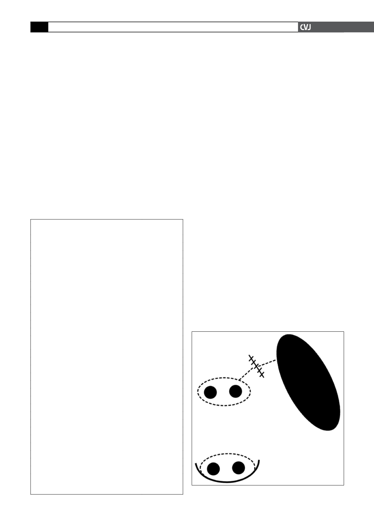
endocardial surface of the left atrium. The left pulmonary veins
were explored and released, epicardial bipolar ablation was done
and both pulmonary veins were isolated as an island. The left
atrial appendage (LAA) was resected from outside. Then lesions
were created at the lines between (1) the left pulmonary vein
lesion and the LAA, and (2) the LAA and the mitral annulus, by
placing one electrode of the catheter on the epicardium and the
other on the endocardium (Fig. 1). The left atrial appendectomy
was sutured from outside following the ablation. Finally, the
mitral valvular procedure was performed.
Follow up
Oral anticoagulation was administered to all patients for three
months. Oral anticoagulants were discontinued on the third
month postoperatively if mitral and/or aortic valve replacement
was not performed, patients were in normal sinus rhythm, or
bi-atrial contraction was present.
TABLE 1. BASELINE DEMOGRAPHICALAND
CLINICAL CHARACTERISTICS
Parameter
n
=
95
Gender, M/F (%)
27/68 (27.8/72.2)
Age, mean
±
SD
57.38
±
11.59
Functional capacity,
n
(%)
NYHA class I–II
11 (11.4)
NYHA class III
62 (65.8)
NYHA class IV
22 (22.8)
Chronic obstructive pulmonary disease,
n
(%)
7 (7.6)
Palpitations,
n
(%)
67 (70.8)
Distribution of structural cardiac pathologies,
n
(%)
Isolated mitral valve disease
63 (66)
Mitral plus aortic valve disease
14 (15)
Mitral valve plus coronary artery disease
14 (15)
Mitral plus aortic valve disease plus coronary
artery disease
4 (4)
Type and aetiology of mitral valve lesions,
n
(%)
Lesion type
Mitral stenosis
22 (23)
Mitral insufficiency
28 (30)
Mixed disease
45 (95)
Aetiology
Rheumatic
70 (74)
Degenerative
17 (18)
Ischaemic
8 (8)
Surgical procedures,
n
(%)
Isolated MVR
59 (62)
MVR plus AVR
14 (14)
MVR plus CABG
6 (7)
Isolated mitral valve repair
4 (5)
Mitral valve repair plus CABG
8 (9)
MVR plus AVR plus CABG
4 (5)
NYHA: NewYork Heart Association; MVR: mitral valve replace-
ment; AVR: aortic valve replacement; CABG: coronary artery bypass
grafting.
Left pulmonary veins
Fig. 1. Lines where the ablation procedure was done.


