CARDIOVASCULAR JOURNAL OF AFRICA • Vol 23, No 6, July 2012
338
AFRICA
patients had gingival and plaque index scores that were rated as
poor.
Abnormalities were detected in the panoramic radiographs
of 84.1% of patients. The most frequent lesion was caries,
present in 56.8% of patients, followed by missing teeth in
54.5%, and impacted teeth in 25% of patients. Retained roots
were present in 22.7% and peri-apical pathology was detected
in 18.1% of patients. Although seven (15.9%) patients had a
normal panoramic radiograph, only one also had both plaque and
gingival index scores that were good.
Thom and Howe reported on their study in which 50 patients,
all with severe heart disease, were examined clinically and
radiologically to ascertain their dental status.
22
Their study differs
from ours in a number of respects. Firstly, they included four
patients with congenital heart disease (ventricular septal defect).
Secondly, they used full-mouth intra-oral peri-apical radiographs,
whereas our study used rotational panoramic radiography.
Thirdly, they decided whether the patients were ‘dentally fit’
and whether they had periodontal disease, although diagnostic
criteria were not given. We studied oral health with particular
reference to oral hygiene. Fourthly, their study included
edentulous patients whereas ours did not have any edentulous
patients. Fifthly, their study was done in a developed country.
Nevertheless, comparisons can be made with our study.
Furthermore, their findings and conclusions are applicable to
developing countries. Thom and Howe reported that 20.5% of
patients were dentally fit.
22
By contrast, only one patient in our
group had a normal panoramic radiograph, a plaque index score
that was rated as good, and a gingival index score that was good
– the latter indicating mild gingival inflammation. Periodontal
disease was present in 46% of their patients. Using only the
clinical examination, 86.5% of our patients had a fair or poor
gingival index. The relative frequency of caries, retained roots
and impacted teeth in their study was 20, 34 and 6%, respectively,
while in our study it was 56.8, 22.7 and 25%, respectively.
These authors divided their patients into one group (
n
= 11)
that visited the dentist regularly (groupA) and another group (
n
=
39) that visited the dentist irregularly (group B). They found that
all clinical and radiographic abnormalities were more frequent
in group B [dentally unfit (36.4 vs 82.1%), retained roots (9.1
vs 41%), caries (0 vs 25.6%), periodontal disease (35 vs 51.3%)
and impacted teeth (0 vs 77%)]. Although statistical tests were
not done in this study, the findings indicate that regular dental
care is beneficial.
Holbrook
et al.
performed a mirror-and-probe examination
of teeth and soft tissue of 100 patients with a cardiac valvular
lesion attending a cardiac clinic; six had a history of infective
endocarditis.
23
They found that only 40.5% of the 42 patients
with teeth could be regarded as having satisfactory dental health;
the remaining patients had either chronic periodontal infection
or an abscess, or both. The dental health of edentulous patients
was also poor – 53.9% had ill-fitting dentures and 28.9% had
diseases of the mouth that could produce bacteraemia.
Smith and Adams published a report on the dental health of
81 at-risk patients attending a cardiology out-patient clinic.
24
This
investigation consisted of a clinical examination and completion
of a questionnaire. Edentulous patients were included in the
study because of several reports in the literature of edentulous
patients suffering from infective endocarditis.
25-27
The criteria
used to classify a patient as dentally unfit were provided.
Forty-eight (59.3%) patients were dentally fit; 25 of these
patients were edentulous. The dentally unfit group comprised 33
(40.7%) patients, of whom four were edentulous. These workers
commented that the prevalence of periodontal disease in their
patients was high; however, no figures were given. The articles
by Thom and Howe, Holbrook and co-workers, and Smith and
Adams, which were based on studies done in the UK, also
indicated that a number of edentulous patients were dentally
unfit.
22-24
In a study of 38 children with congenital heart disease, dental
examination revealed dental caries in 39% of the children.
28
In another study, 42.4% of 170 children with congenital heart
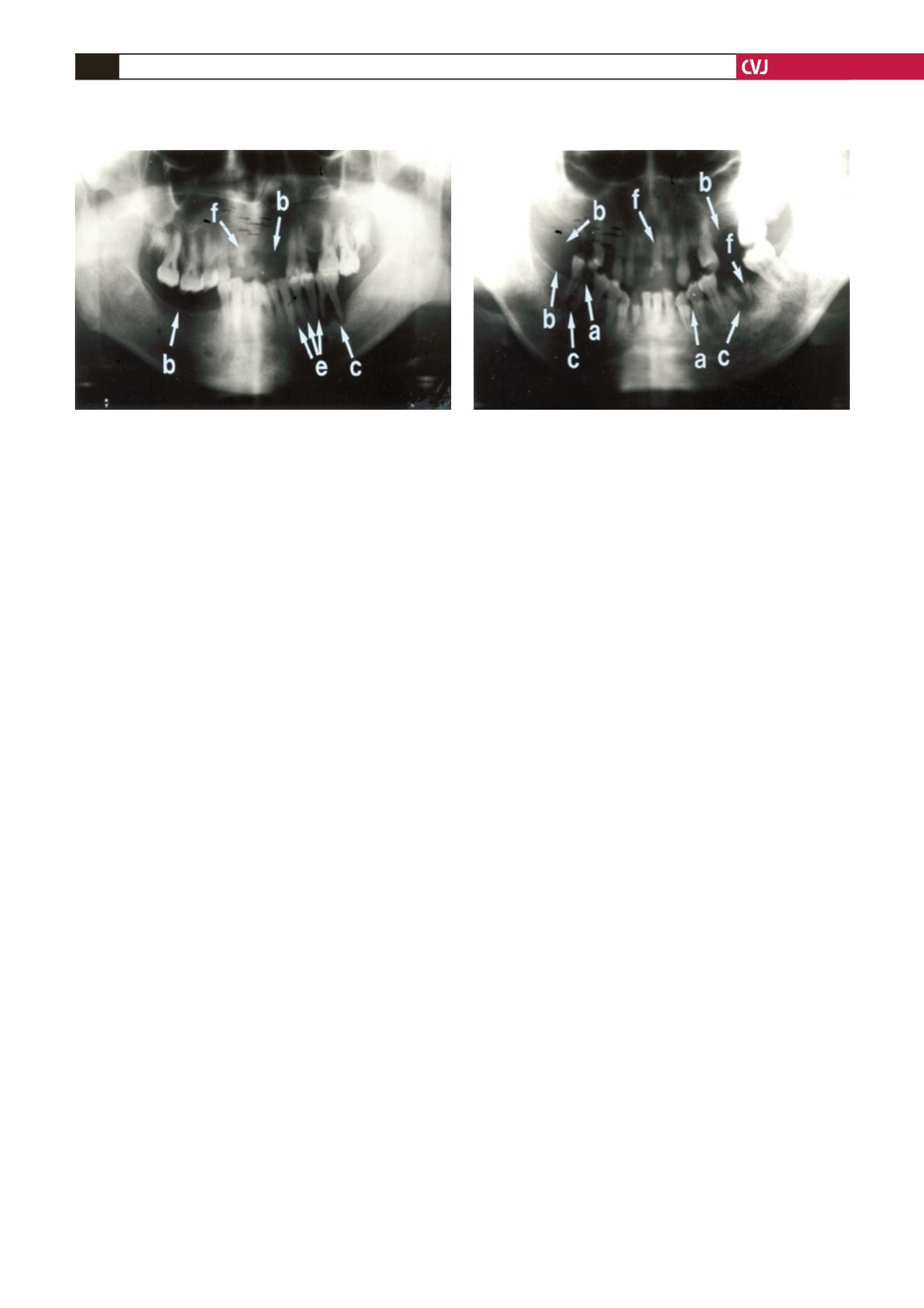
Fig. 3. Panoramic radiograph showing more abnormali-
ties than Fig. 2. This radiograph shows edentulous areas
(i.e. loss of teeth) involving the right mandibular molar
region and the left maxillary incisor and canine regions
(b). A retained root of the right maxillary canine tooth is
also present (f). Interdental bone loss is seen between
the left mandibular premolar and molar teeth (e). A
peri-apical radiolucency is associated with the left first
mandibular molar tooth (c). The overall condition of the
teeth of this patient is poor.
Fig. 4. Panoramic radiograph showing many abnormali-
ties. This radiograph shows multiple retained roots (f)
and missing teeth (b) in both mandibular and maxillary
arches. The right mandibular first molar tooth shows a
large carious lesion with loss of crown enamel (a). The
left mandibular canine teeth and the left mandibular first
premolar tooth show caries interproximally (a). Peri-
apical radiolucencies are noted in relation to the right
and left mandibular molar teeth (c). The overall condition
of the teeth of this patient is extremely poor.


