CARDIOVASCULAR JOURNAL OF AFRICA • Volume 25, No 3, May/June 2014
e6
AFRICA
haemorrhage and contiguous spread of the infection to the
neighbouring spine or paravertebral soft tissue. This leads to
soft tissue swelling and compression of the vertebral neural root,
resulting in neurological deficits.
3
The clinical approach for diagnosingMAA includes laboratory
tests and radiological imaging. On laboratory examination,
leukocytosis is found in 64 to 71% of patients,
4
and inflammatory
markers, including CRP, as well as the erythrocyte sedimentation
rate (ESR) are usually elevated. Blood cultures may be negative
in 25 to 50% of patients,
5
and are therefore not sufficient to
exclude an infected aneurysm.
An MRI scan may be the most sensitive and specific imaging
technique for detecting infected soft tissue, bone destruction
and abscess formation in the early stage of infection. However,
a disadvantage is its longer duration and inability to easily
distinguish rupture of the pseudolumen. Enhanced CT is also
useful for diagnosing MAA, and for revealing the pseudolumen,
soft tissue swelling, as well as abscess formation, and is of
shorter duration. It is optimal for studying infected aneurysms,
particularly if the patient’s haemodynamic status is unstable.
3
Management of MAA using traditional open aortic repair is
a major surgical challenge in diseased patients because of the
high associated rates of mortality and morbidity (13–40%).
1,6,7
Traditional open aortic repair has a complicated management
strategy, including surgical resection and debridement of the
infected aorta and surrounding tissues, the use of muscle flaps
or omentum to cover the infected field, and either an
in situ
interposition graft or an extra-anatomical bypass.
In the report by Chen
et al
.,
3
there were six patients with MAA
who presented with radiculopathy (Table 1). Most of the pathogens
were
Salmonella
. The patients were treated with aggressive surgical
debridement and aneurysm resection. Four patients survived a
two-year follow up; two were walking with walking aids.
3
EVAR provides a less invasive approach, lower surgical risk
compared to conventional open repair, and more favourable
result. It also has the advantage of avoiding a large incision, full
heparinisation and extracorporeal circulation, and minimises
the need for blood transfusion. However, complex anatomy, an
unresected infective aneurysm, and remnant paravertebral soft
tissue are the major problems of EVAR. Recurrent infection
may result in disastrous consequences, so further antibiotic
therapy and adjunct procedures, such as surgical debridement or
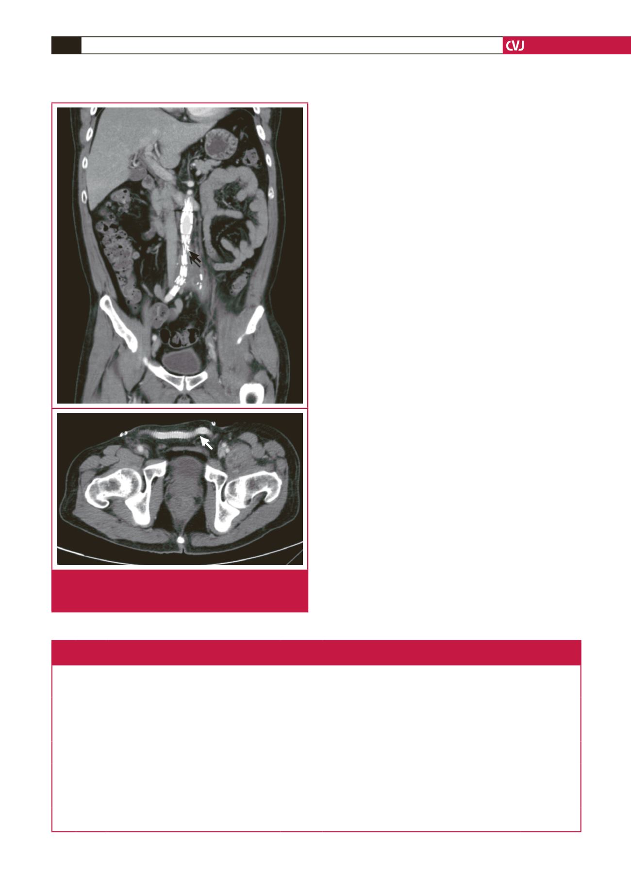
Fig. 2.
An aortic-to-right iliac stent graft was placed (black
arrows), and a femoral-to-femoral bypass with a pros-
thetic graft was performed (white arrow).
Table 1. Summary of clinical details, surgical methods and outcome of six patients with MAA
presenting with radiculopathy (from Chen
et al
.
3
)
Case
No
Age/
gender Clinical symptoms
Symptom
duration
Lesion site
of radicu-
lopathy Pathogen
Surgical methods
Outcome
1 60/M Back pain with fever
8 weeks T12–L1
Salmonella
Open surgical aneurysm resection +
debridement + vertebral reconstruction
Survived
2 50/M Back pain with fever
2 weeks L3–L4
Salmonella
Open surgical aneurysm resection +
debridement
Survived
3 79/M Back pain with fever, abdominal
pulsating mass, lower limb weakness
1 week L3–L4
Salmonella
Mycobacterium
Open surgical aneurysm resection +
debridement + vertebral reconstruction
Survived
4 72/M Back pain with fever, abdominal
pulsating mass
3 weeks L3–L4
Salmonella
Open surgical aneurysm resection +
debridement
Survived
5 81/M Back pain with fever, abdominal pain 4 weeks L5
Streptococcus
None
Died (aneurysm
rupture)
6 59/F Back pain with fever, papaplegia
1 week T7
Staphylococcus
None
Died (septic
shock)


