CARDIOVASCULAR JOURNAL OF AFRICA • Vol 23, No 9, October 2012
e8
AFRICA
segment of the left anterior descending artery (LAD) (Fig. 1).
Transthoracic echocardiography (TTE), performed in the
coronary intensive care unit, revealed inferior wall hypokinesia
with an ejection fraction of 55%, mechanical mitral valve area
of 2 cm
2
and 8 mmHg mean gradient. No obvious thrombus
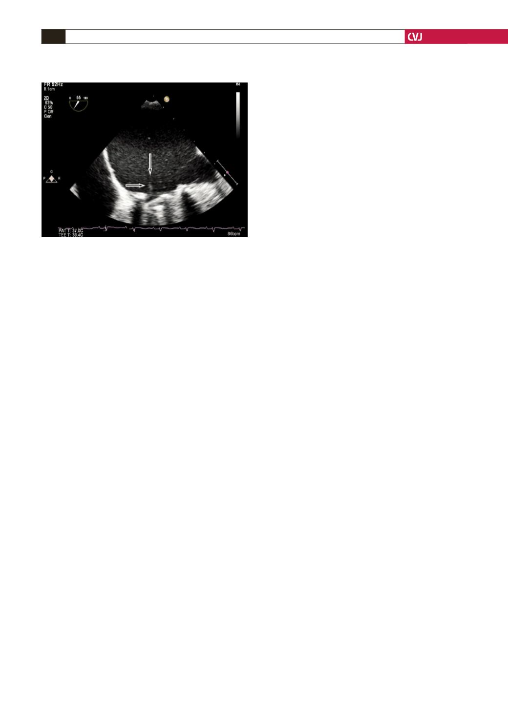
was seen. For futher evaluation, TEE was performed and a
non-obstructive prosthetic mitral valve thrombosis was seen
(
Fig. 2).
Low dose (25 mg) and slow infusion (6 hours) of tissue
plasminogen activator (t-PA) was administered. Control coronary
angiography was normal without any residual thrombus in the
LAD. A day after thrombolysis, TEE revealed complete lysis of
the prosthetic valve thrombus.
Discussion
Patients with a prosthetic heart valve and acute coronary
syndrome who present with non-ST elevation myocardial
infarction are a rare subgroup, more likely to be elderly, with risk
factors for atherosclerosis. It is reported that the pathogenesis of
acute coronary syndrome is commonly coronary atherosclerotic
disease rather than prosthetic heart valve-derived emboli.
10
Coronary embolism is a rare entity with prosthetic heart valves,
and even rarer with mitral prostheses. In the literature, most
cases were caused by aortic valve prostheses with obvious
thrombus burden.
Optimal management of these situations remains controversial,
although surgery is usually favoured.
7,11
The use of thrombolysis
may be limited by the increased risk of dislodgement of the
embolus, distal embolisation and haemorrhagic complications.
Additionally, rapid infusion of thrombolytic agents has also
been associated with higher complication rates.
8
By determining
the lowest effective infusion dosage and duration, the risk of
thromboembolism and haemorrahage can be minimised.
Our patient presentedwithacute inferior transmuralmyocardial
infarction in Killip class I. She had been asymptomatic for three
years following mitral valve replacement and was in normal
sinus rhythm throughout. At presentation, there was no clinical
evidence of PVT on TTE but TEE revealed a non-obstructive
trombus on the prosthetic mitral valve. Although it is reported
that the pathogenesis of acute coronary syndromes is usually
coronary atherosclerotic disease rather than PVT-derived
emboli,
10
our case suggests the possibility of a small thrombus
burden, which embolised to the distal part of the LAD, with
no prosthetic valve dysfunction or secondary emboli to other
organs.
While present data suggest the administration of 100 mg
t-PA with the usual protocol for myocardial infarction, due to
its risk of complications, the decision was made to administer
low-dose (25 mg), prolonged infusion (6 hours) of t-PA to
achieve gradual lysis at the lowest effective dose to minimise
thromboembolic and haemorrhagic complications. We recently
presented the efficacy and safety of this treatment modality in
patients with PVT.
12
Inadequate anticoagulation is the most likely
complicating factor.
Conclusion
We report the occurence of a coronary embolism, presenting as an
acute ST-elevation myocardial infarction, in a young patient with
a mitral valve replacement. It was found to be a non-obstructive
prosthetic valve thrombosis. She was successfully treated with
low-dose, prolonged infusion of tPA.
References
1.
Cannegieter SC, Rosendaal FR, Brier E. Thromboembolic and bleed-
ing complications in patients with mechanical heart valve prosthesis.
Circulation
1994;
89
: 635–641.
2.
Sun JCJ, Davidson MJ, Lamy A, Eikelboom JW. Antithrombotic
management of patients with prosthetic heart valves: current evidence
and future trends.
Lancet
2009;
374
: 565–576.
3.
Salem DN, Stein PD, Ahmad AA, Bussey HI, Horstkotte D, Miller N,
et al
.
The Antithrombotic therapy in valvular heart disease – native
and prosthetic: Seventh ACCP Conference on Antithrombotic and
Thrombolytic Therapy.
Chest
2004;
126
: 457–482.
4.
Burchfiel CM, Hammermeister KE, Krause-Steinrauf H, Sethi GK,
Henderson WG, Crawford MH,
et al
.
Left atrial dimension and risk of
systemic embolism in patients with prosthetic heart valve.
J Am Coll
Cardiol
1990;
45
: 32–41.
5.
Cáceres-Lóriga FM, Pérez-López H, Santos-Gracia J, Morlans-
Hernandez K. Prosthetic heart valve thrombosis: pathogenesis, diagno-
sis and management.
Int J Cardiol
2006;
110
: 1–6.
6.
Horstkotte D, Burkhardt D. Prosthetic valve thrombosis.
J Heart Valve
Dis
1995;
4
: 141–153.
7.
Lengyel M, Horstkotte D, Völler H, Mistiaen WP; Working Group
Infection, Thrombosis, Embolism and Bleeding of the Society for Heart
Valve Disease. Recommendations for the management of prosthetic
valve thrombosis.
J Heart Valve Dis
2005;
14
(5): 567–575.
8.
Özkan M, Kaymaz C, Kirma C, Sönmez K, Özdemir N, Balkanay M,
et al
.
Intravenous thrombolytic treatment of mechanical prosthetic valve
thrombosis: a study using serial transesophageal echocardiography.
J
Am Coll Cardiol
2000;
35
(7): 1881–1889.
9.
Charles RG, Epstein EJ, Holt S, Coulshed N. Coronary embolism in
valvular heart disease.
Q J Med
1982;
51
: 147–161.
10.
Iakobishvili Z, Eisen A, Porter A, Cohen N, Abramson E, Mager A,
et
al
.
Acute coronary syndromes in patients with prosthetic heart valves –
A case-series.
Acute Cardiac Care
2008;
10
(3): 148–151.
11.
Tong AT, Roudaut R, Ozkan M, Sagie A, Shahid MS, Pontes Júnior SC,
et al.
prosthetic valve thrombolysis-role of transesophageal echocar-
diography (PRO-TEE) registry investigators.
J Am Coll Cardiol
2004;
43
(1): 77–84.
12.
Biteker M, Duran NE, Gündüz S, Kaya H, Kaynak E, Çevik C,
et al
.
Comparing different intravenous thrombolytic treatment regimens in
patients with prosthetic heart valve thrombosis under the guidance of
serial transesophageal echocardiography: A 15-year study in a single
center (TROIA Trial).
Circulation
2008;
118
: 932.
Fig. 2. Transoesophageal echocardiographic imaging
showing a mobile, non-obstructive thrombus on the
mechanical prosthetic mitral valve (arrows).


