CARDIOVASCULAR JOURNAL OF AFRICA • Vol 22, No 4, July/August 2011
AFRICA
205
Cardiogenic shock due to dynamic left ventricular
outflow tract obstruction of acute myocardial infarction:
an under-diagnosed complication
CY KARABAY, G KOCABAY, A KALAYCI, H TANBOGA, M MERT, C KIRMA
Abstract
We report on a patient who developed cardiogenic shock
caused by dynamic left ventricular outflow tract (LVOT)
obstruction following percutaneous coronary intervention
for anteroseptal acute myocardial infarction.
Keywords:
dynamic left ventricular outflow tract obstruction,
myocardial infarction, shock, esmolol
Submitted 18/5/10, accepted 30/6/10
Published online: 10/1/11
Cardiovasc J Afr
2010;
22
: 205–206
DOI: 10.5830/CVJA–2010–051
Acute dynamic left ventricular outflow tract (LVOT) obstruction
has been reported as a complication of myocardial infarction.
1
The incidence of this obstruction is unclear and it may well be
under-diagnosed. This mechanical complication of myocardial
infarction should be ruled out since the treatment of this condi-
tion differs completely from that of acute coronary syndrome.
Case report
A 60-year-old male patient with no known disorder was admit-
ted to the emergency deparment of our hospital for chest pain of
three hours’ duration. He also complained of angina following
effort over the last month. Physical examination showed that his
blood pressure was 140/80 mmHg and pulse rate was rhythmical
and 90 beats/minute. His other body systems were normal.
The ECG investigation showed anteroseptal myocardial
infarction. He was taken to the catetherisation laboratory and
angiography revealed that he had 100% obstruction in the
proximal portion of the left anterior descending artery (LAD). A
3.0
×
20-mm bare-metal stent was placed in the lesioned vessel
following a 2.0
×
20-mm balloon application. Thrombolysis in
myocardial ınfarction (TIMI) 3 flow was achieved.
The patient developed dyspnoea one hour after the stent was
placed. He became pale and hypothermic; his blood pressure
was 70/40 mmHg and pulse rate 130 beats/minute. His physical
Kartal Kosuyolu Yuksek Ihtisas Heart Education and
Research Hospital, Department of Cardiology, Istanbul,
Turkey
CY KARABAY, MD
G KOCABAY, MD,
A KALAYCI, MD
H TANBOGA, MD
C KIRMA, MD
Kayseri Educational and Research Hospital, Department of
Endocrinology and Metabolism, Kayseri, Turkey.
M MERT, MD
examination revealed a new holosystolic ejection murmur, grade
3/6 in the apical area, with radiation to the axillary artery.
Transthoracic echocardiography was performed to exclude
possible mechanical complications. Echocardiography revealed
anterior anteroseptal hypokinesia, systolic anterior motion of the
anterior leaflet of the mitral valve and mild mitral regurgitation.
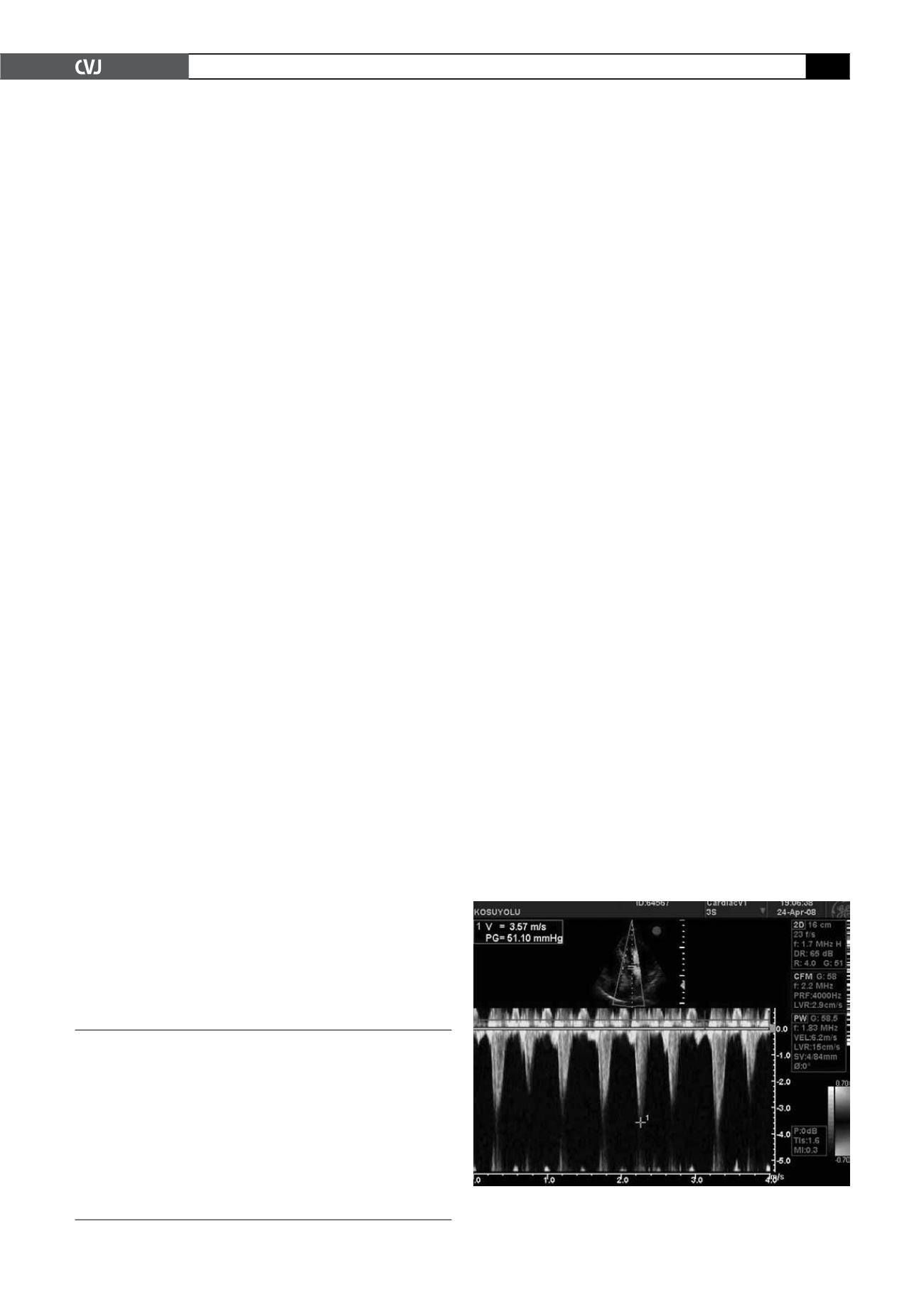
The ejection fraction was 35%. Doppler examination revealed the
LVOT gradient to be a maximum gradient of 51 mmHg (Fig. 1).
Under close monitoring, intravenous fluids and esmolol were
initiated. With reduction of the heart rate to below 70 beats/
min, the murmur and other symptoms disappeared. After two
hours, echocardiography was repeated and showed no LVOT
obstruction. The patient was subsequently discharged in a stable
condition.
Discussion
The reason for a dynamic LVOT obstruction in the presence of
acute coronary syndrome is usually due to compensated hyper-
dynamic basal wall motion in patients with antero-apical infarc-
tion. Hyperdynamic basal wall motion causes decreased LVOT
cross-sectional area.
3
The treatment of this situation is different from that for
myocardial infarction. The use of vasodilators (nitrates), inotrop-
ic agents (dopamine, epinephrine, dobutamine), intra-aortic
balloon pump and volume depletion are contraindicated. Utilising
alpha-agonists, beta-blockers and intravenous fluids reduces the
gradient.
1
Beta-blockers may help by decreasing hyperkinesis
of the basal segments and decreasing the left ventricular gradi-
ent and hypotension.
4
In the present case, since the patient was
hypotensive, we administered esmolol because of its rapid onset
and very short duration of action.
Fig. 1. Doppler examination showed the LVOT gradient to
be a maximum of 51 mmHg.


