CARDIOVASCULAR JOURNAL OF AFRICA • Vol 22, No 6, November/December 2011
e2
AFRICA
× craniocaudal). In addition, a subpleural bulla approximately
2 cm in diameter was observed in the anterior apex of the left
upper lobe, and marked compressive atelectasis of the left lower
lung was noted. No additional mediastinal, hilar, airway or lung
parenchyma abnormalities were identified.
As the mass was symptomatic and the diagnosis remained in
question, the patient was scheduled for operative exploration.
The mass, thought to be a pericardial cyst, was removed via an
open thoracotomy.
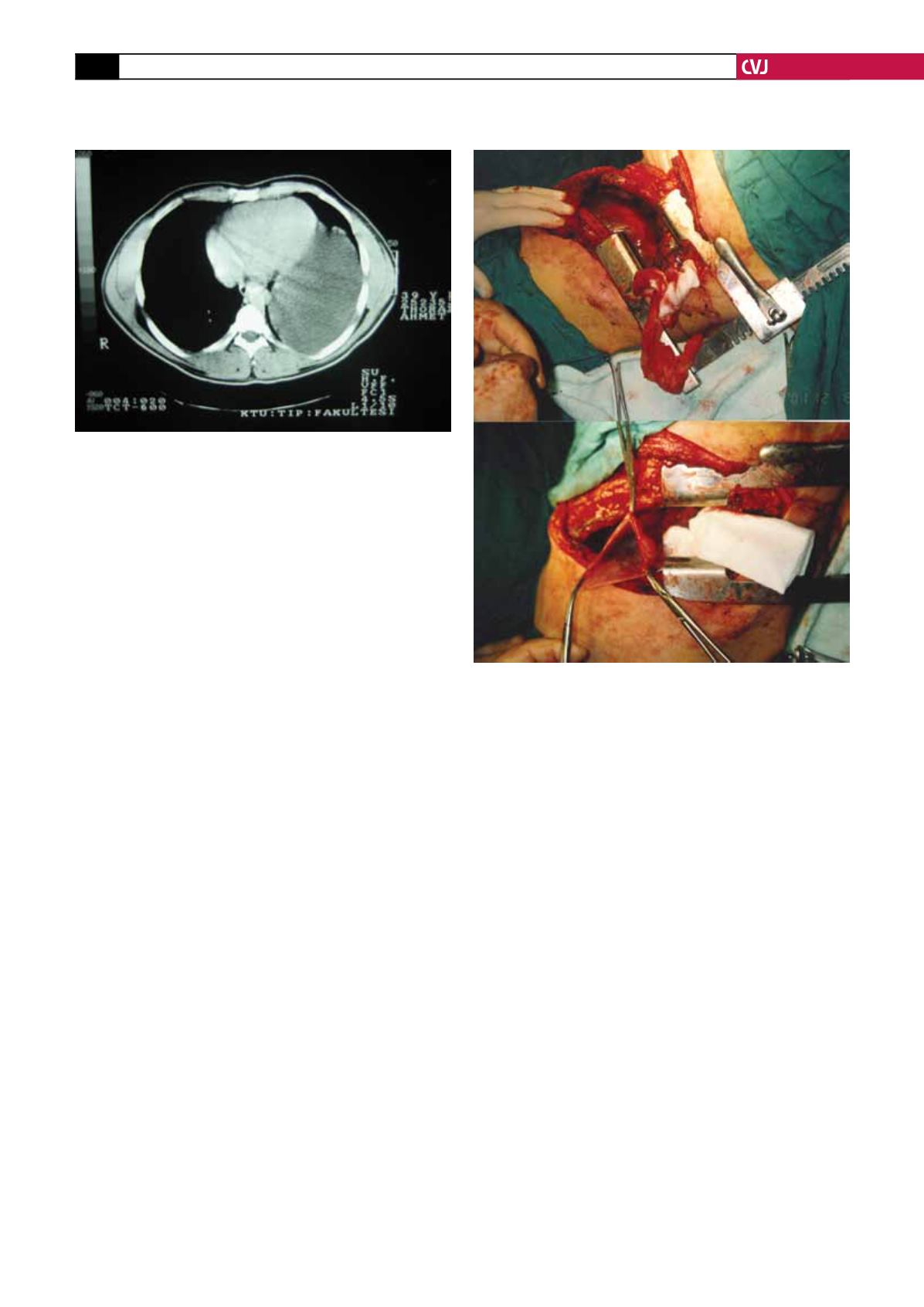
After a left-sided thoracotomy, 2 000 ml of serosanguinous
fluid were aspirated from the pericardial cyst sac, with complete
excision of the mediastinal mass, measuring 22
×
15
×
17 cm
(Fig. 3). The cyst was full of golden-yellow liquid and was adher-
ent to the lung and phrenic and anterior vagus nerves laterally.
The pericardial cyst was connected to the inferior pericardial
surface by a thin, mobile, vascular pedicle, which was ligated
when the mass was resected.
The mass was bright and soft. It was on the left side of the
diaphragm, and exerted pressure on the left side of the heart
and pericardium and on the lower lobe of the left lung, causing
compressive atelectasis. The atelectic lung was expanded manu-
ally. The 2
×
2-cm bulla in the left upper lobe of the lung was
sutured. The procedure was finished successfully in the usual
manner after closed drainage (water-sealed drainage) of the
thoracic cavity.
Histological examination confirmed the diagnosis of a peri-
cardial cyst (fibrovascular cyst wall) with no evidence of malig-
nancy or tissue other than pericardium. Cultures of both the
cystic and pericardial liquids were negative. Notably, no giant
cells, granulomata, acid-fast bacilli or fungi were identified.
No postoperative complications were noted. The patient had
an unremarkable recovery and was discharged on postoperative
day six.
Discussion
Pericardial cysts are benign intrathoracic lesions that occur
in one in every 100 000 persons and constitute 7% of all
mediastinal tumours.
5
Pericardial cysts can be suspected if the
patient complains of non-specific chest pain, dyspnoea, cough
or epigastric fullness. Although complications are uncommon,
unexpected life-threatening events have been reported, such as
acute cardiac tamponade,
6
and sudden death.
7
No cases of malig-
nant degeneration have been reported. Spontaneous resolution
of pericardial cysts is unusual, although two cases of presumed
spontaneous resolution have been reported.
8
The cysts range in size from 2–3 cm, up to a maximum of 28
cm reported by Braude
et al
.
9
In our case, the pericardial cyst
measured 22
×
15
×
17 cm. As the patient had seen many differ-
ent doctors, including a psychiatrist, with various complaints, the
diagnosis was rather belated.
Of all pericardial cysts, 70–75% are located at the right cardio-
phrenic angle, 22% at the left, and the rest are in the posterior or
anterior superior mediastinum.
3
In our case, the pericardial cyst
occurred in the left cardiophrenic area, to which it was attached
by a pedicle over the fat tissue on the left side of the pericardium.
Solid tumours should be considered in the differential diagno-
sis, including angiomas, lipomas, neurogenic tumours, sarcomas,
lymphomas, bronchogenic carcinomas, metastases, granuloma-
tous lesions, and abscesses, along with interstitial bronchogenic
cysts, lymphangiomas, diaphragmatic hernias, aneurysms of the
heart or great vessels, and other diseases.
10
Once a pericardial cyst is suspected on the chest X-ray,
thoracic CT with intravenous contrast is commonly used to
confirm the diagnosis. However, the diagnosis of a pericardial
cyst using CT can be challenging, and the exact location cannot
always be ascertained.
11
In our case, although CT was conducted
in another hospital, a diaphragmatic hernia was diagnosed, and
the patient was transferred to our clinic for surgery.
Fig. 2. Chest computed tomography shows a unilocular
cystic mass at the left cardiophrenic angle.
Fig. 3. Intra-operative view of intact pericardial cyst (top)
and pericardial cyst wall after fluid drainage (bottom).


