CARDIOVASCULAR JOURNAL OF AFRICA • Vol 23, No 7, August 2012
e2
AFRICA
a mean of 53 mmHg. Cardiac magnetic resonance imaging
(MRI) showed diffuse circumferential mid-wall and epicardial
enhancement in the late phase following gadolinium contrast,
in keeping with extensive myocardial fibrosis. There was no
evidence of active inflammation, oedema or infarction (Fig. 2).
His condition was extremely debilitating and his prognosis
was poor. After extensive discussions between the patient, his
cardiologist, the heart failure team and the cardiac surgeon,
it was felt the only route to improve his quality of life was to
undergo valve surgery despite the increased risk of mortality.
Aortic valve replacement with mitral and tricuspid valve
repair was undertaken after the patient recovered from his acute
admission. Intra-operative transoesophageal echocardiography
confirmed the presence of torrential AR, with severe MR and TR
due to marked annular dilatation. Severe biventricular dilatation
with extremely poor function was also confirmed. A size 28-mm
Edwards Physio mitral and a size 32-mm rigid tricuspid ring
were implanted. The aortic valve was replaced with a 19-mm
Edwards Magna-Ease valve. He made a good post-operative
recovery, and his symptoms have since improved dramatically.
The excised native aortic valve leaflets were thickened
with retracted cusps (Fig. 3A). Microscopically, there was a
fibrosed inflamed aortic valve with dense fibroelastic tissue,
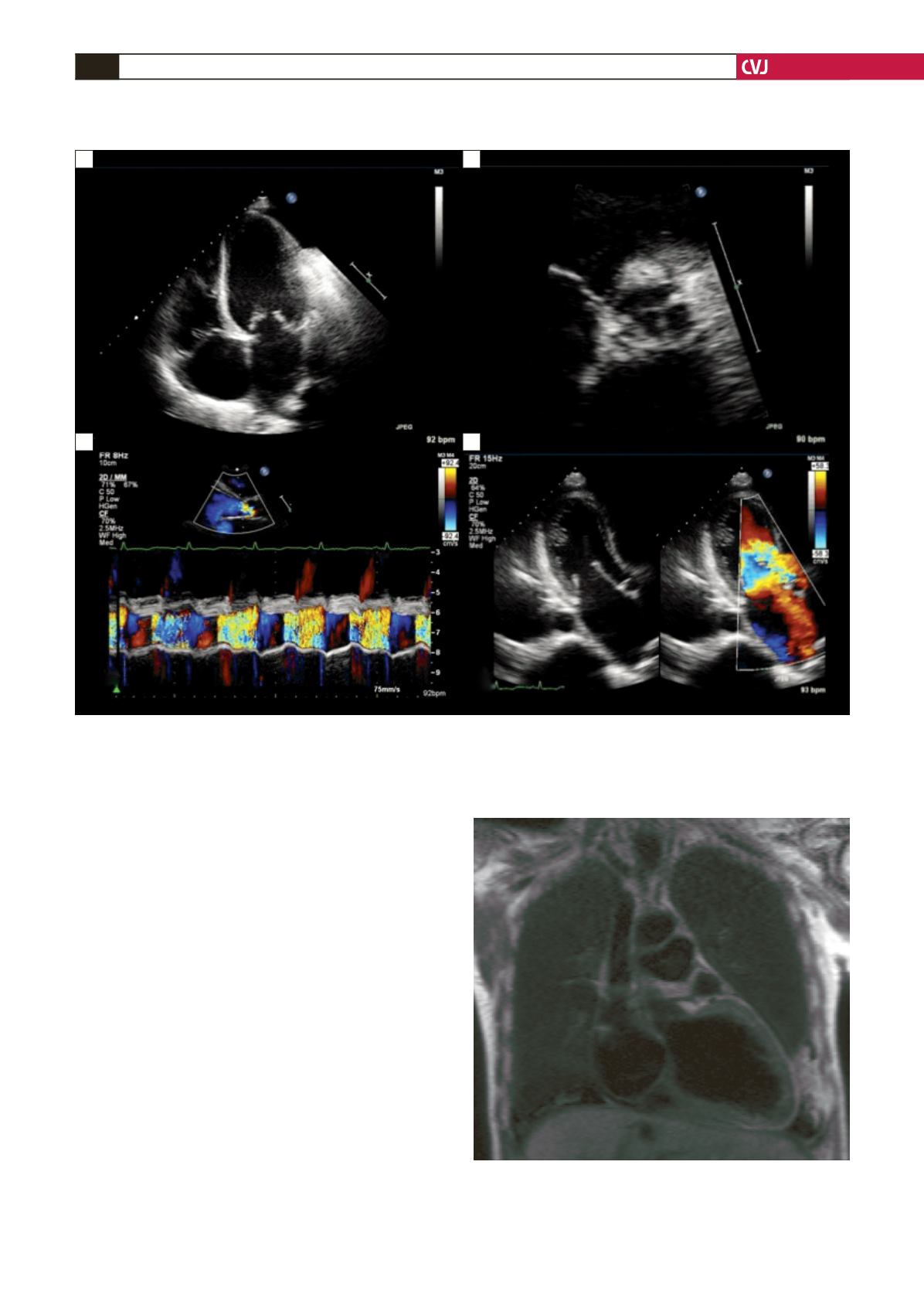
Fig. 1. Echocardiography: four-chamber view showing bi-atrial and bi-ventricular dilatation and thickening of the mitral
valve and tricuspid valve leaflets (A), short-axis view showing thickening of the aortic valve leaflets with a mal-coaptation
defect between the left coronary cusp and non-coronary cusp (B), M-mode with colour flow across the aortic valve show-
ing a non-dilated aortic valve root with severe broad aortic regurgitation (C), and three-chamber view with colour flow
showing severe eccentric mitral regurgitation (D).
Fig. 2. Cardiac MRI in late phase after gadolinium contrast
showing diffuse, circumferential mid-wall and epicardial
enhancement consistent with extensive myocardial fibro-
sis.
A
C
B
D


