CARDIOVASCULAR JOURNAL OF AFRICA • Vol 23, No 7, August 2012
AFRICA
e3
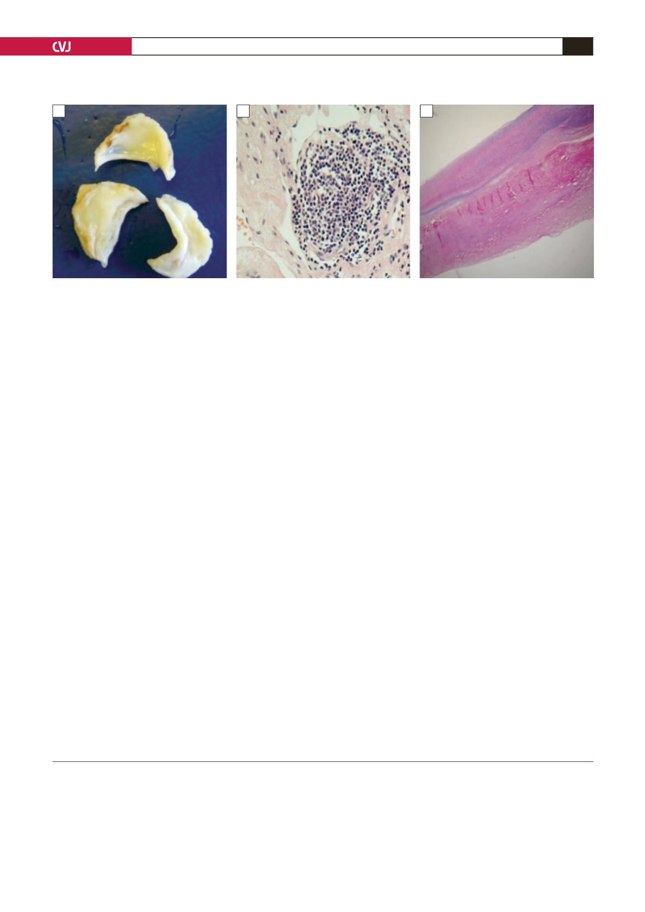
neovascularisation and focal lymphocyte infiltration (Fig. 3B,
C). There was no evidence of sarcoidosis in the valve or
surrounding lymph node. No rheumatoid nodules, granuloma or
calcification were seen. These findings supported the diagnosis
of valvular heart disease secondary to RA.
Post-operative echocardiography showed a well-seated
prosthetic aortic valve with no AR, mild MR, no TR, and no
pericardial collection. At one month, the LVEDD was improved
to 6.0 cm. Echocardiography 10 months post surgery showed
a well functioning aortic prosthetic valve with a significant
reduction in LVEDD from 6.5 cm to 4.6 cm. Mitral and tricuspid
valve function was improved, with no regurgitation seen 10
months post repair.
Discussion
Pre-operative non-invasive cardiac imaging supported a diagnosis
of severe valvular heart disease due to RA, and aggressive steps
were taken to treat his heart failure. Valve thickening was
seen on echocardiography, which is a distinctive feature that
provides evidence of valve involvement in RA.
5
Myocardial
fibrosis demonstrated on cardiac MRI gave further support to
this diagnosis. The pattern of delayed enhancement helped to
distinguish from other causes of myocardial fibrosis, including
sarcoidosis and ischaemic cardiomyopathy.
Most patients with RA-associated valvular heart disease
have mild valvular insufficiency due to a slowly progressive
granulomatous valvulitis.
2
Rapidly progressive, severe
Fig. 3. Macroscopic view of the aortic valve showing thickened and retracted valve cusps (A). Histology slides showing
lymphocyte infiltration and new vessel formation (B), and fibroelastic tissue (C).
non-granulomatous valve disease has also been described in
RA.
2
This was the diagnosis in our patient.
Conclusion
This case report highlights potential difficulties in the
management of patients with severe heart failure due to
rheumatoid arthritis. Early surgical intervention is often the best
treatment option in this setting, as severe left ventricular failure
is unlikely to respond to medical treatment alone and valve
replacement surgery can improve left ventricular function and
prolong survival.
6
References
1.
Lebowitz W. The heart in rheumatoid arthritis.
Ann Intern Med
1063;
58
: 102–123.
2.
Libby P, Bonow RO, Zipes DP, Mann DL. Rheumatic diseases and
the cardiovascular system. In:
Braunwald’s Heart Disease
. 8th edn.
Philadelphia: Saunders Elsevier 2007; 2094–2096.
3.
Voskuyl AE. The heart and cardiovascular manifestations in rheumatoid
arthritis.
Rheumatology
2006;
45
: iv4–iv7.
4.
Kaplan MJ. Cardiovascular complications of rheumatoid arthritis –
assessment, prevention and treatment.
Rheum Dis Clin North Am
2010;
36
(2): 405–426.
5.
Roldan CA, DeLong C, Qualls CR, Crawford MH. Characterization of
valvular heart disease in rheumatoid arthritis by transoesophageal echo-
cardiography and clinical correlates.
Am J Cardiol
2007;
100
: 496–502.
6.
Levine AJ, Dimitri WR, Bonser RS. Aortic regurgitation in rheumatoid
arthritis necessitating aortic valve replacement.
Eur J Cardiothorac
Surg
1999; 213–214.
A
B
C


