


 52 / 64

CARDIOVASCULAR JOURNAL OF AFRICA • Volume 27, No 1, January/February 2016
50
AFRICA
laminae. There was no evidence of vasculitis or atherosclerosis.
The overall features were in keeping with a diagnosis of
intimomedial mucoid degeneration.
The patient’s renal function and blood pressure improved
postoperatively. However on the third day after admission
his haemoglobin dropped and abdominal distension was
noted. A repeat CTA indicated haemorrhage around the auto-
transplanted kidney with possible leakage of the abdominal
aneurysm. The patient received an emergent hybrid repair of the
extensive thoraco-abdominal aneurysm. The procedure involved
debranching of the coeliac artery, superior mesenteric artery
(SMA) and right renal artery, with extensive stent-graft repair
using four overlapping aortic stent grafts.
Prior to the stent-grafting, the coeliac artery and SMA were
revascularised with a bifurcated prosthetic graft, using the
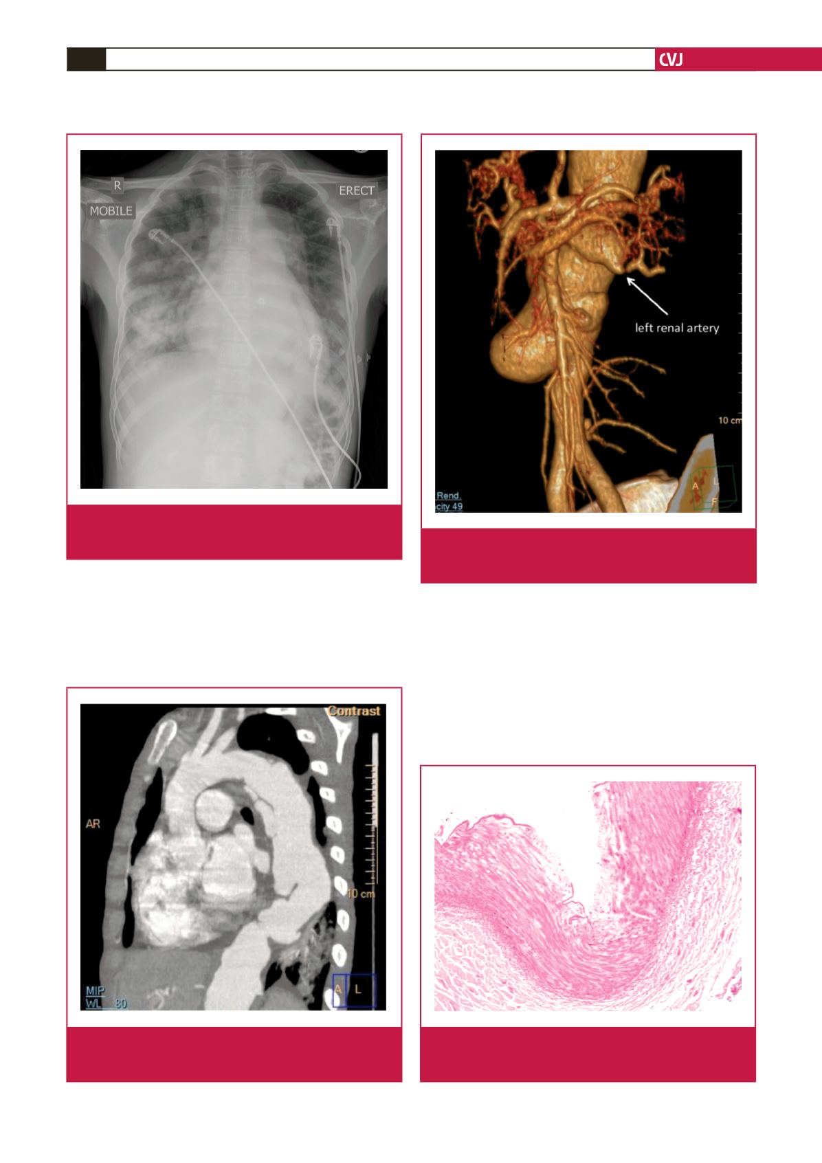
Fig. 3.
Volume-rendered CT reconstruction shows the aneu-
rysmal abdominal aorta as well as aneurysmal origin
of the left renal artery with focal stenosis.
Fig. 2.
Oblique sagittal MIP reconstruction shows a diffusely
aneurysmal descending thoracic aorta with complex
multi-level dissection flaps.
Fig. 4.
Mild intimal thickening with disruption of the internal
elastic lamina is shown on this haematoxylin and eosin
stain (200
×
magnification).
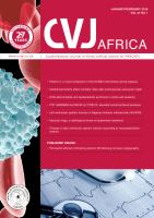
Fig. 1.
Admission AP frontal chest radiograph demonstrating
extensive pulmonary oedema as well as widening of
the superior mediastinum.