CARDIOVASCULAR JOURNAL OF AFRICA • Vol 21, No 5, September/October 2010
260
AFRICA
In the Ch group, the density of positive cells was high, and
HO-1 was expressed in the endothelial and foam cells in plaques.
The expression rate of HO-1 was significantly higher than in the
C group (40.98
±
2.47 vs 19.02
±
1.28%,
p
<
0.01). In the Hm
group, the density of positive cells was very high, and HO-1 was
widely expressed in the endothelial and smooth muscle cells. The
expression rate of HO-1 was significantly higher than in the Ch
group (90.84
±
6.42 vs 40.98
±
2.47%,
p
<
0.01).
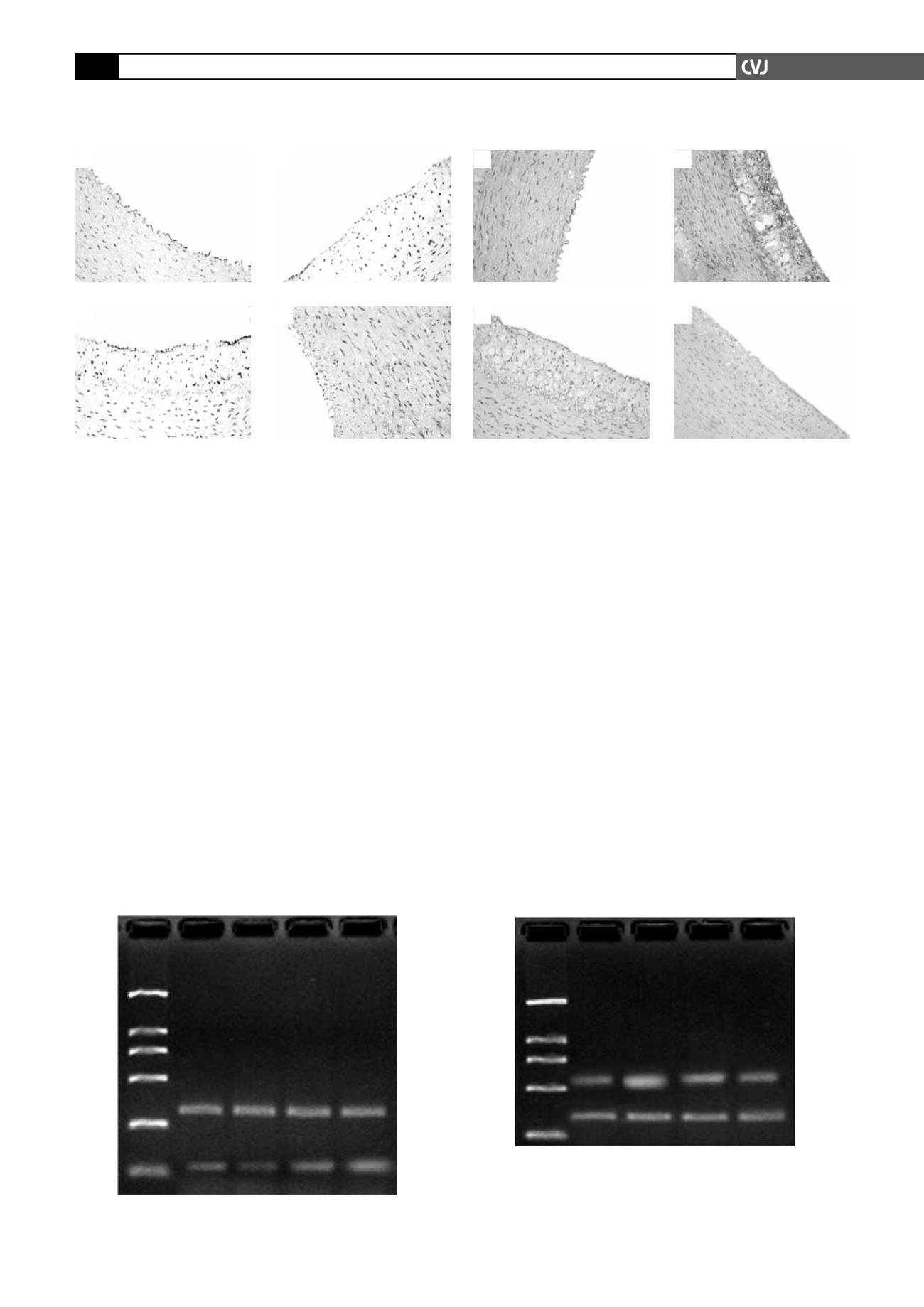
As shown in Figs 3 and 4, the immunohistochemical staining
and PCR of ET-1 showed that there was no obvious ET-1 expres-
sion in the aortic wall in the C group (16.08
±
1.30%). The Zn
and Ch groups showed strongly positive ET-1 expression (74.16
±
4.28, 59.28
±
3.42, vs 16.08
±
1.30%,
p
<
0.01), and the Hm
group showed weakly positive expression (23.71
±
1.49 vs 16.08
±
1.38%,
p
<
0.01).
mRNA expressions of HO-1 and ET-1 in aortic tissue
Compared with the C group, HO-1 mRNA expression in both the
Ch and Hm groups was significantly increased (1.27
±
0.16, 1.61
±
0.22 vs 1.06
±
0.12, all
p
<
0.01), especially in the Hm group
with a 1.2-fold increase. HO-1 mRNA expression was signifi-
cantly decreased in the Zn group (0.84
±
0.15 vs 1.06
±
0.12
p
<
0.01). Compared with the Ch group, HO-1 mRNA expression
was significantly increased in the Hm group (1.61
±
0.22 vs 1.27
±
0.16,
p
<
0.01).
The RT-PCR result showed that ET-1 mRNA expression in
the Hm, Ch and Zn groups was significantly higher than in
the C group (1.07
±
0.09, 1.59
±
0.16, 2.11
±
0.25 vs 0.75
±
0.15, all
p
<
0.01). Compared with the Ch group, ET-1 mRNA
expression was significantly decreased in the Hm group (1.07
±
0.09 vs 1.59
±
0.16,
p
<
0.01), whereas ET-1 mRNA expres-
Fig. 2. Immunohistochemical detection in the C (A),
Zn (B), Ch (C) and Hm (D) groups of the buffy lamel-
lar particles of HO-1 present in the cytoplasm and cell
membrane. HO-1 is mainly expressed by the endothe-
lial, foam and smooth muscle cells of the tunica–intima
(magnification 200 X).
A
B
C
D
Fig. 3. Immunohistochemical detection in the C (A), Zn
(B), Ch (C) and Hm (D) groups of ET-1. Buffy lamellar
particles of ET-1 were located in the endothelial cells,
foam cells and smooth muscle cells of the tunica–intima
of the aorta (magnification 200 X).
A
B
C
D
M C Zn Ch
Hn
bp
2000
1000
750
500
250
100
b
-actin
312
HD-1
118
M C Zn Ch
Hn
bp
2000
1000
750
500
250
ET-1
543
b
-actin
312
Fig. 4. Levels of HO-1 (left) and ET-1 (right) mRNA expression in the aorta. M: marker, C, Zn, Ch and Hm groups.


