
CARDIOVASCULAR JOURNAL OF AFRICA • Vol 24, No 2, March 2013
30
AFRICA
M-mode and two-dimensional echocardiography
Simultaneous M-mode and two-dimensional echocardiography
was performed. M-mode recordings were made in the parasternal
long-axis (PLAX) position during apnea with the cursor at
the level of the chordae tendineae and papillary muscles.
PLAX, parasternal short-axis (PSAX), apical four-chamber
(A4C) and apical two-chamber (A2C) views were taken in
ciné-loop format and recorded on DVD discs in both DICOM
and MPEGVUE for subsequent evaluation by the independent
team. An echocardiography report was written according to the
laboratory’s protocol and handed to the patient.
Severity of the valvular lesion was classified according
to the ACC/AHA.
13
Left ventricular and left atrial dilatation
were defined as left ventricular diastolic diameter and left
atrial diameter more than 57 and 40 mm, respectively.
13
Left
ventricular systolic dysfunction was defined as ejection fraction
less than 55%.
13
Pulmonary artery systolic pressure (PASP) was
estimated from the peak velocity of the tricuspid regurgitation jet
plus the estimated right atrial pressure. Patients with PASP
≥
30
mmHg were classified into mild (
<
50 mmHg), moderate (50–79
mmHg) and severe (
≥
80 mmHg) pulmonary hypertension.
13
Statistical analysis
Data were captured into EPI-DATA (version 3.1), cleaned and
then exported to Stata version 10 for analysis. Continuous
variables were summarised as mean (
±
standard deviation)
and median (inter-quartile range), and presented in the tables.
Categorical data were analysed using frequency and percentages,
and results are presented in frequency tables and bar charts. Test
of significance (
p
-value) was determined using the chi-square
test. A
p
-value of less than 0.05 was considered significant.
Results
We screened over a period of eight months, 156 patients who
were suspected clinically of having RHD, using the echo
machine. Twenty-six patients were excluded for the following
reasons: probable/possible RHD (13 cases), normal echo
findings (two cases), congenital heart disease (six cases), dilated
cardiomyopathy (four cases) and cor pulmonale (one case);
130 patients who were confirmed to have definite RHD were
recruited and entered in the data analysis (Fig.1).
Table 1 shows the demographic characteristics of the 130
newly diagnosed cases of RHD. Overall, females (72.3%)
predominated, with a younger median age of males than females
(24 vs 33 years). The majority of the study population’s highest
education level was primary school (total: 46.2%; male: 52.8%;
female: 43.6%), while 10% (male: 8.3%; female: 10.6%) were
illiterate. Unemployment rate was as high as 64.6% (male:
52.8%; female: 69.2%) and 32.3% (male: 44.4%; female:
27.7%) lived in temporary houses.
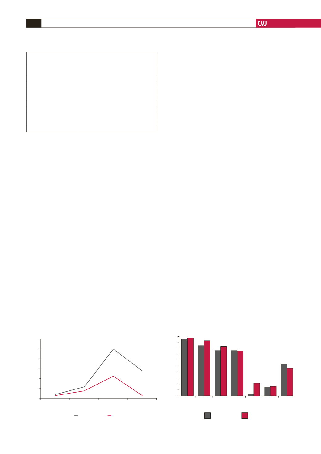
The age distribution of newly diagnosed RHD patients
showed a peak in the young adult age group (20–39 years). The
disease was lowest in the age group
<
12 years (5.4% of RHD
cases), increased in the 12–19-year group (15.4%), peaked at
20–39 years (55.4%), followed by a declining number of case
presentations in the age group 40–65 years (23.8%). The pattern
of case presentation according to age was similar for males and
females (Fig. 2).
Fig. 3 shows the frequencies of symptoms with which the
study participants presented. Palpitations were the commonest
symptom (95.4%), followed by fatigue (89.2%) and dyspnoea
(75%). Other symptoms included chest pain (74.6%), syncope
(15.4%) and oedema (14.6%). There were no gender-specific
statistical differences in most of the symptoms, except females
reported more syncope than males (20.2 vs 2.8%) and more
males presented with severe heart failure than females.
Table 2 shows the frequency distribution of rheumatic valve
lesions by age group. Isolated or multiple valve lesions were
observed in the spectrum of RHD. There were eight types of
valvular lesions detected according to the valve affected. Pure
mitral regurgitation (MR) was the most prevalent lesion (55
cases, 42.3%), followed by MR + aortic regurgitation (AR) (36
TABLE 1. SOCIO-ECONOMIC DATA OF NEWLY DIAGNOSED
RHEUMATIC HEART DISEASE PATIENTS (
n
=
130)
All
(
n
=
130)
Females
(
n
=
94)
Males
(
n
=
36)
Gender distribution (%)
100
72.3
27.69
Median age (years)
29.5
33
24
Educational level
none,
n
(%)
13 (10)
10(10.64)
3(8.33)
primary,
n
(%)
60 (46.15)
41 (43.62)
19 (52.78)
secondary,
n
(%)
42 (32.31)
31 (32.98)
11 (30.56)
college/university,
n
(%)
16 (12.31)
8 (8.51)
8 (22.22)
No formal employment
84 (64.62)
65 (69.15)
19 (52.78)
Temporary housing
42 (32.31)
26 (27.66)
16 (44.44)
Fig. 2. Age distribution of newly diagnosed RHD.
60
50
40
30
20
10
0
<
12
12–19
20–39
40–65
females
males
Number of cases
Years
Fig. 3. Frequncy of symptoms in newly dignosed patients.
100
90
80
70
60
50
40
30
20
10
0
palpita-
tions
fatigue dysp-
noea
chest
pain
syncope oedema NYHA
(III/IV)
males
females
p
= 0.81
p
= 0.23
p
= 0.31
p
= 0.90
p
= 0.007
p
= 0.35
p
= 0.47
Symptoms
Percentage


