CARDIOVASCULAR JOURNAL OF AFRICA • Vol 24, No 3, April 2013
AFRICA
e5
right pulmonary vein was draining into a stenosed inferior vena
cava, just before it entered the RA. A gradient of 15 mmHg at
this junction was demonstrated.
At cardiac catheterisation, there was a step-up in the high IVC
saturation. Severe pulmonary hypertension of 80/22 mmHg was
demonstrated. Angiography demonstrated a hypertrophied and
dilated RV, a large MPA and an absent RPA (Fig. 3). PAPVC
of the right pulmonary vein draining into the IVC was also
noted (Fig. 4). The stenosed IVC was clearly demonstrated
before its entry into the RA (Fig. 5). There was a hemi-azygous
continuation which drained into the superior vena cava (SVC)
and the RA (Fig. 5).
A systemic collateral from the descending aorta supplying the
lower lobe of the right lung was visible during the venous phase
of the pulmonary angiogram. A diagnosis of scimitar syndrome
with an absent RPA and an obstructed IVC was made. In view
of the anal atresia, recto-vaginal fistula, hemi-vertebrae with
scoliosis, right thumb hypoplasia and scimitar syndrome, the
criteria for diagnosis of the VACTERL association were fulfilled.
Due to the absent RPA and right lung hypoplasia, surgical
repair of the PAPVC and re-routing of the anomalous right
pulmonary vein into the left atrium (LA) were considered of little
haemodynamic benefit. Right pneumonectomy was deemed the
best surgical option, but could not be undertaken in early infancy.
The patient initially improved with the administration of
anti-failure treatment (furosemide and digoxin) and could be
discharged from hospital. Unfortunately, she succumbed a few
weeks later to a lower respiratory tract infection while at home
and an autopsy could not be performed.
Discussion
Scimitar syndrome is a rare form of PAPVC involving the
right lung. There is associated hypoplasia of the right lung
and RPA with a right mediastinal shift creating dextroposition
of the heart. The RPA may be completely absent,
3-5
as was the
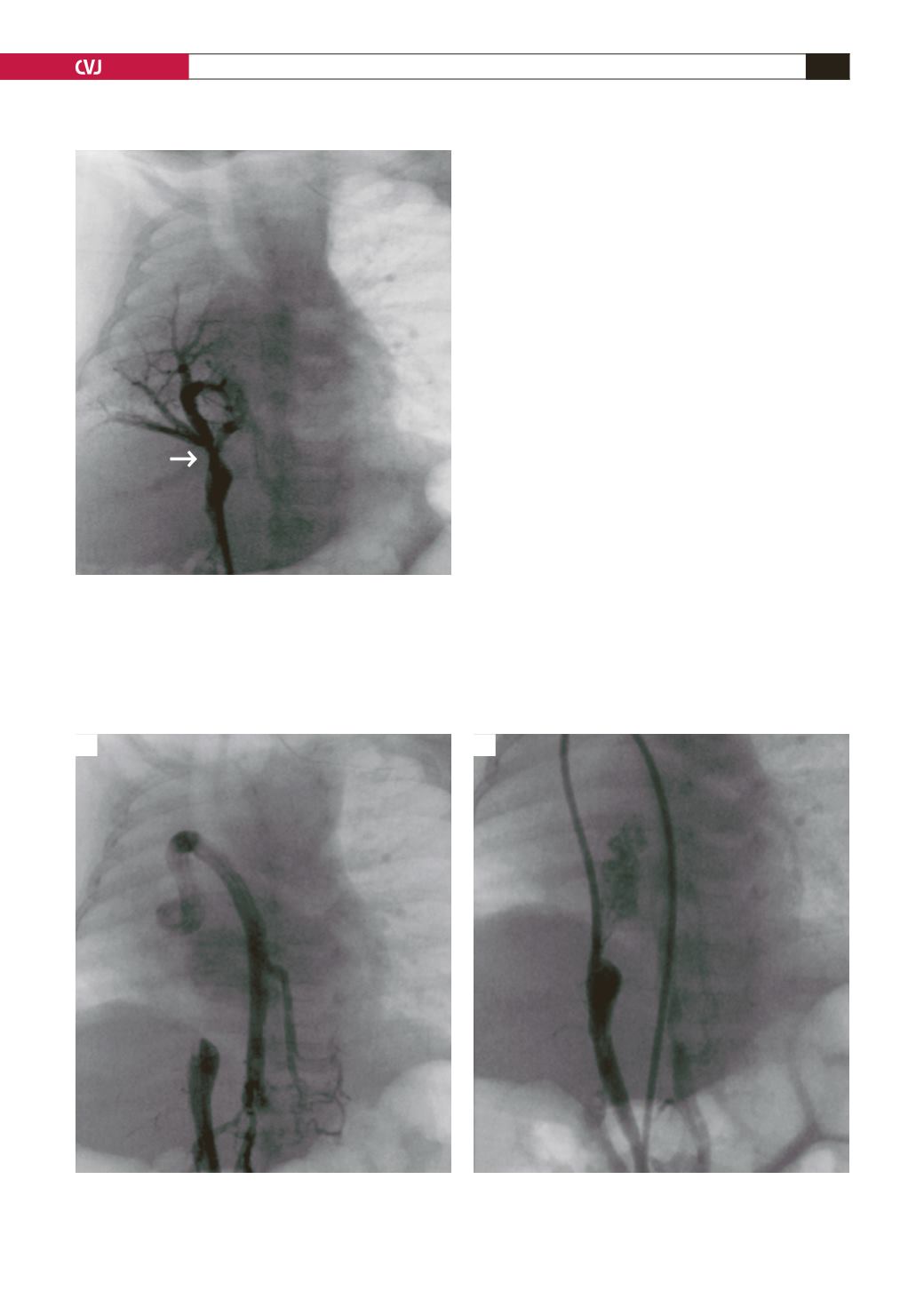
Fig. 4. Abnormal right pulmonary vein draining into the
stenosed inferior vena cava (arrow).
SVC
HZ
IVC
IVC
Fig. 5. (A) Obstructed IVC with hemi-azygous continuation draining into the SVC. (B) Catheter advanced from the
femoral vein into the hemi-azygous continuation, SVC, RA, and into the IVC (IVC, inferior vena cava; SVC, superior
vena cava; HZ, hemi-zygous, RA, right atrium).
A
B


