


 71 / 78

CARDIOVASCULAR JOURNAL OF AFRICA • Volume 28, No 6, November/December 2017
AFRICA
e1
Case Report
Efficacy of cardiac magnetic resonance imaging in a
sub-aortic aneurysm case
Ruchika Meel, Richard Nethononda, Ferande Peters, Mohammed Essop
Abstract
Sub-aortic (SA) aneurysms are a rare entity of variable aetiol-
ogy. We report the first case of a SA aneurysm assessed using
cardiac magnetic resonance imaging (MRI). A 33-year-old
female with human immunodeficiency virus and on highly
active antiretroviral treatment presented with syncope and
dyspnoea. Clinical examination suggested moderate to severe
aortic regurgitation (AR) confirmed by transthoracic and
transoesophageal echocardiograms. However, echocardiog-
raphy was suboptimal in defining the precise mechanism and
severity of AR. A cardiac MRI was done to elucidate the aeti-
ology, severity and mechanism of regurgitation. It confirmed
the presence of a SA aneurysm below the left coronary cusp
and its retraction, resulting in an eccentric AR jet. An assess-
ment of moderate AR, based on regurgitant volume, was
made. Furthermore, the anatomical relationships of the aneu-
rysm were clearly defined. Cardiac MRI allowed comprehen-
sive assessment of this SA aneurysm.
Keywords:
cardiac magnetic resonance imaging, sub-aortic aneu-
rysm, aortic regurgitation
Submitted 25/1/17, accepted 1/5/17
Published online 29/6/17
Cardiovasc J Afr
2017;
28
: e1–e3
www.cvja.co.zaDOI: 10.5830/CVJA-2017-027
Sub-aortic (SA) aneurysms are a rare entity with variable
aetiology. Most cases are congenital and result from a defect
between the ventricular wall and valvular annuli.
1
Earlier reports
were mostly from Africa. Between 1957 and 1993, only 22 cases
had been reported.
2
Since then, isolated reports on various
aspects of this rare condition have been published. There
have been no reports in the literature on adult patients, using
cardiac magnetic resonance imaging (MRI), to investigate SA
aneurysms.
Case report
The patient was a 33-year-old human immunodeficiency virus
(HIV)-positive woman on highly active antiretroviral treatment
(HAART), with a current CD4 count of 1 000 cells/
µ
l. She was
referred from a peripheral hospital, with a history of a single
syncopal episode. She also admitted to a two-week history of
progressive dyspnoea and fatigue (New York Heart Association
functional class II). No further relevant past medical or family
history was obtained.
On examination, the blood pressure was 102/52 mmHg with
a pulse of 106 beats/min. No dysmorphic features were noted.
She had large volume and collapsing peripheral arterial pulses,
with a wide pulse pressure (50 mmHg). The apex beat was in
the fifth intercostal space and displaced slightly to the left of
the midclavicular line. The second heart sound was loud. There
was a grade 3/4 early decrescendo diastolic murmur in the left
parasternal border, characteristic of aortic regurgitation (AR).
There were no peripheral stigmata of infective endocarditis.
She had been treated with diuretics and there were no signs of
congestive cardiac failure.
An electrocardiogram showed left ventricular (LV)
hypertrophy with strain pattern in the lateral leads and left atrial
(LA) enlargement. The chest X-ray was normal. The blood
count was normal and the serology for syphilis and connective
tissue disease was negative.
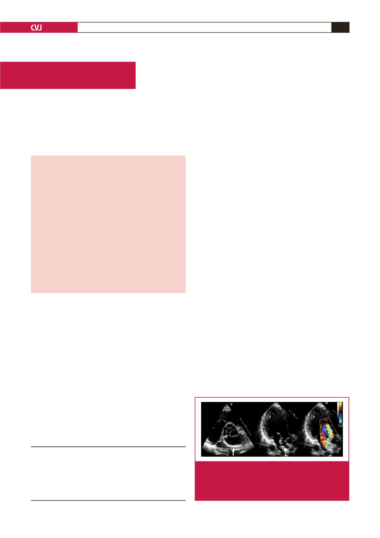
A transthoracic echocardiogram (TTE) revealed a dilated
LV with an ejection fraction of 56% and moderate-to-severe
eccentric aortic regurgitation secondary to leaflet malcoaptation
(Fig. 1). There was compression of the LA by an outpouching
with calcified walls adjacent to the aortic root, the exact origin
and location of which was difficult to define on TTE. There was
flow into and out of this structure in diastole.
Division of Cardiology, Chris Hani Baragwanath
Academic Hospital and University of the Witwatersrand,
Johannesburg, South Africa
Ruchika Meel, PhD,
ruchikameel@gmail.comRichard Nethononda, DPhil
Ferande Peters, MD
Mohammed Essop, MD
Fig. 1.
Parasternal short-axis view of the sub-aortic (SA) aneu-
rysm (white arrow, left). Apical three-chamber views
depicting the SA aneurysm (white arrow, middle), and
an eccentric aortic regurgitation jet on colour flow, with
flow into the SA aneurysm (right).