


 64 / 68

CARDIOVASCULAR JOURNAL OF AFRICA • Volume 30, No 3, May/June 2019
e4
AFRICA
attachment to the inter-atrial septum near the anterior leaflet
of the mitral valve. The myxoma was obstructing the outflow
of the pulmonary veins to the left atrium. It was also prolapsing
onto the left ventricle through the mitral valve in diastole. An
additional myxoma (40 × 25 mm) was found near the left inferior
pulmonary vein (Fig. 2).
The small myxoma with surrounding normal tissue was
completelyexcised.The largeonewas removedwithapproximately
1 cm of the inter-atrial septum and the atrial septal defect was
closed with a bovine pericardial patch. After irrigation of the
atria and ventricle with saline solution to eliminate any tumour
fragments, the left atrium was closed. Cardiopulmonary bypass
was stopped with sinus rhythm following defibrillation. The
duration of cardiopulmonary bypass was 100 minutes.
Case 2: The mother had a history of two cardiac operations
for left atrial myxoma in 2008 and 2012, at the same time
as her daughter. She had fatigue, exertional dyspnoea and
hyperpigmentation on her skin. TTE of the mother revealed
a pedunculated mass (23.9 × 20 mm) that was attached to the
inter-atrial septum (Fig. 3).
The mother had a third cardiac operation with a median
sternotomy accompanied by moderate hypothermia and
cardiopulmonary bypass via the internal jugular vein, femoral
vein and femoral arterial cannulation. An incision was made in
the right atrium and the trans-septal approach was used.
A large jelly-like pedunculated mass (1.96 × 2.39 mm) was
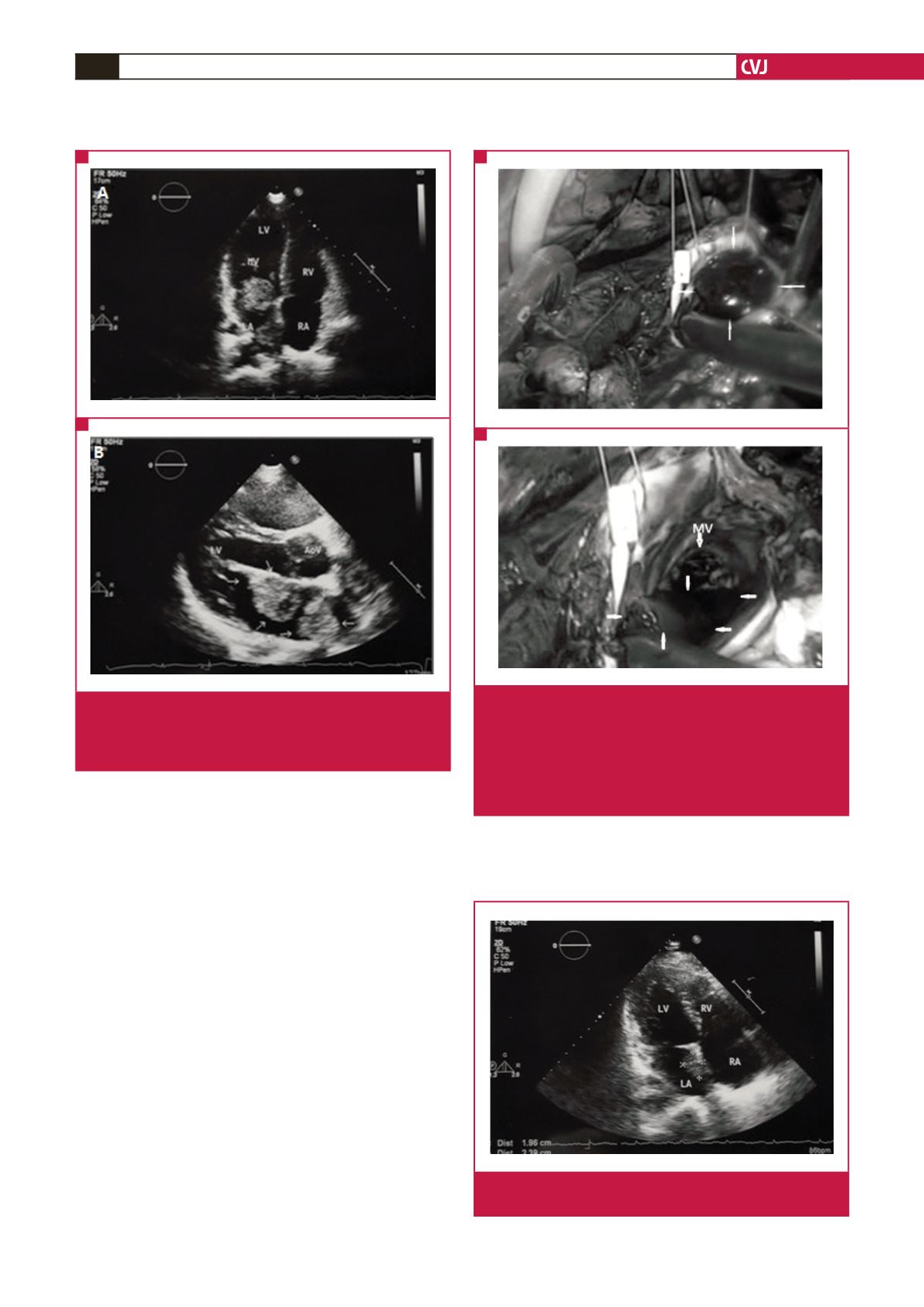
Fig. 1.
Transthoracic echocardiography of case 1. (A) A large
myxoma is bulging through the mitral valve. (B) The
large myxoma is attached to the inter-atrial septum and
a second one is located on the left pulmonary veins.
A
B
Fig. 2.
Macroscopic appearance of the atrial myxomas of
case 1 on a surgical field. (A) The large myxoma
attached to the inter-atrial septum is bulging into the
left ventricle through the mitral valve. (B) The second
left atrial myxoma (its edges are demarcated by
arrows) is located on the orifice of the left pulmonary
veins.
A
B
Fig. 3.
Transthoracic echocardiography of case 2 showing the
left atrial myxoma attached to the inter-atrial septum.