
CARDIOVASCULAR JOURNAL OF AFRICA • Volume 32, No 4, July/August 2021
226
AFRICA
behaves as a benign and asymptomatic lesion and is easily
confused with other intracardiac masses, cysts, thrombus or
abscess.
Under this condition, an association between the CCMA
and a medical history of hypertension, chronic renal failure or
haemodialysis, and abnormal calcium metabolism should be
checked.
3
No related medical history was found in our patient,
which was one of the reasons for the misdiagnosis.
Another reason that led to misdiagnosis may have been the
relatively small size and atypical imaging of the mass. CCMA
is usually large, round, calcified and enveloped in a echolucent
core, typically located at the base of the posterior leaflet,
and can be misdiagnosed as a cardiac tumour or abscess on
echocardiography. The posterior leaflet becomes thickened,
stretched and arched over the mass.
Secondary to these anatomical changes, mitral valve
dysfunction (either stenosis or regurgitation) can be detected.
Since the mass was mimicking a benign cardiac tumour that
had increased in size and could not be differentiated from the
degenerative valves and with significant valvular dysfunction,
surgery was performed on the patient.
MCEmay provide much more information about the location,
border and perfusion of the CCMA, however no report has been
published on the details. According to its pathological findings,
the authors concluded that a ring-enhancement mass with a
no-perfusion core should be detected by MCE. However, the
present case did not seem to fit the characteristics.
The CCMA had a small volume and edge with abundant
neovascularisation, which was misleading since the mass was
vascular. Another potential contributing factor was the ‘bleeding’
effect, which means the blood of the surrounding left ventricular
cavity was moving into the regions of interest as the mass was
rapidly oscillating in and out of the imaging plane. We therefore
suggest that when MCE is used to evaluate the vascularity of an
intracardiac mass, operators need to be aware of these potential
pitfalls.
Cardiac computed tomography (CT) and magnetic resonance
imaging (MRI) also can be helpful in confirming or establishing
diagnosis. Non-contrast CT showed a large calcified mass
at the base of the posterior mitral annulus, extending to the
adjacent mitral valve and myocardium.
6
On contrast-enhanced
CT, the central part appeared less hypodense due to the caseous
toothpaste-like material contained within the denser calcified
peripheral rim.
8-10
MRI has shown low signal on both T1- and
T2-weighted images due to calcification but was inferior in
showing the calcification directly.
9
Surgical intervention is not only the definitive treatment to
remove the potential obstacle of obstruction or embolisation
the mass brings about, but also a way to accurate diagnosis
and therapy. Therefore our patient had the mass resected and
the valve replaced. She is now in a good general condition
and undergoing out-patient care with follow up and further
management.
Conclusion
CCMA is an exceedingly rare valvular lesion with an excellent
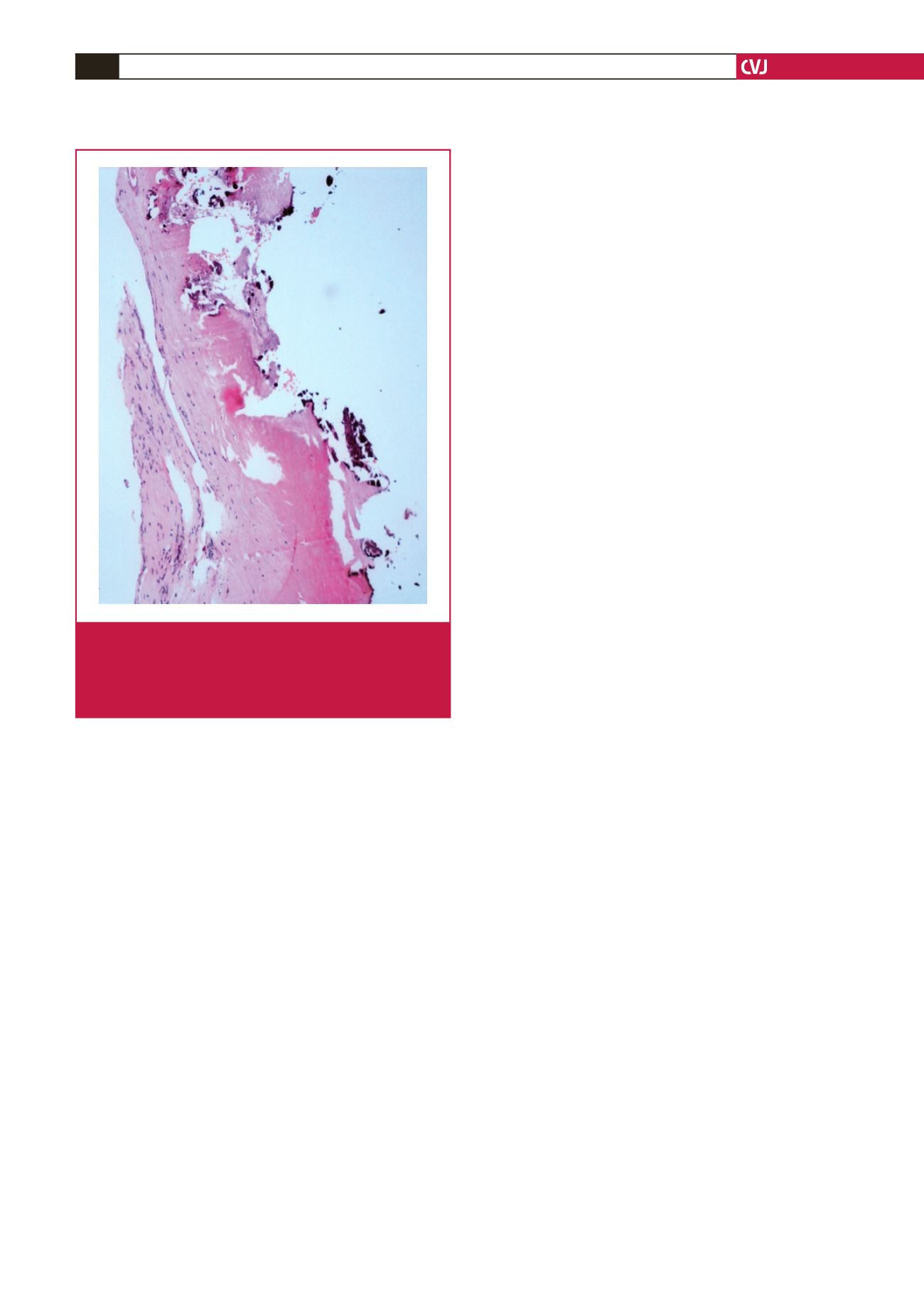
outcome following complete surgical removal. Histopathological
findings of an amorphous, acellular, basophilic and calcific
structure, with a chronic inflammatory response, is the gold
standard for diagnosis. Although it was difficult to differentiate
from other cardiac masses via echocardiography, diagnosis could
be made based on combined imaging modalities. This incidental
lesion may be encountered in clinical practice now and then, and
cardiac imaging interpretation experts should be familiar with it
in order to avoid misdiagnosis.
This study was supported by grants from the National Natural Science
Foundation, People’s Republic of China (No. 81371571); Zhejiang Provincial
Natural Science Foundation, People’s Republic of China (No. LY13H180008);
Scientific Research Fund of Zhejiang Provincial Education Department (No.
Y200907825); Health and Family Planning Commission of Zhejiang Province
(2016KYA078); and the platform key programme of Health and Family
Planning Commission of Zhejiang Province (grant 2015ZDA013).
References
1.
Deluca G, Correale M, Leva R, Del Salvatore B, Gramenzi S, Di Biase
M. The incidence and clinical course of caseous calcification of the
mitral annulus.
J Am Soc Echocardiogr
2008;
21
: 828–833.
2.
Martinez-de-Alegria A, Rubio-Alvarez J, Baleato-Gonzalez S. Caseous
calcification of the mitral annulus: a rare cause of intracardiac mass.
Case Rep Radiol
2012;
201
2: 596962.
3.
Shintaro K, Sunao W, Kohei A, Manabu Y, Joji I,
et al.
Caseous calci-
fication of mitral annulus.
Cardiovasc Diagn Tber
2013;
3
(2): 108–110.
4.
Plank F, AI-Hassan D, Nguyen G,
et al
. Caseous calcification of the
mitral annulus.
Cardiovasc Diagn Tber
2013;
3
(2): E1–3
Fig. 3. Pathological examination of the resected tissue
revealed hyperplasia of the tissue (mitral valve) with
myxoid and calcific degeneration, and it also had
some scattered chronic inflammatory cell infiltrate into
the tissue.


