CARDIOVASCULAR JOURNAL OF AFRICA • Vol 23, No 4, May 2012
AFRICA
e9
abdominal cavity revealed a large, inoperable tumour of the
terminal ileum. A carcinoid tumour was diagnosed from the
histological specimens. The first liver metastases were detected
in 1997.
After the first symptoms of CS (flushing, diarrhoea, oedema
of the ankles) emerged in 2002, somatostatine analogue therapy
was introduced. This led to good control of the CS symptoms,
and the urinary levels of 5-hydroxyindoleacetic acid (5-HIAA),
a byproduct of serotonin degradation used for the detection
and follow up of patients with carcinoid tumours, significantly
decreased.
In 2007 the patient started to complain of fatigue and
dyspnoea on exertion. An echocardiographic examination (Aloka
SD 4000 and Philips i22) revealed pathognomonic signs of CHD
– severe thickening, retraction, shortening and almost complete
immobilisation of the tricuspid valve leaflets (Fig. 1a) with
severe tricuspid regurgitation (Fig. 1b), mild tricuspid stenosis,
pulmonary valve stenosis and severe dilatation and trabeculation
of the right ventricle (Fig. 1c).
The continuous-wave Doppler signal analysis showed a
characteristic dagger-shaped signal of tricuspid regurgitation with
a maximal velocity of 240 cm/s, demonstrating rapid decline in
pressure difference between the right ventricle and right atrium
(Fig. 1d). As a result of volume overload of the right ventricle,
diastolic flattening of the interventricular septum was noticed.
No pericardial effusions were detected but hyperechogenicity
of the pericardium was noted and MR imaging of the heart was
performed to exclude pericardial involvement. Cardiac MRI
was performed using the 3.0-Tesla MRI system (Magnetom
Trio, Siemens Medical Solutions, Erlangen, Germany) with
a 12-element cardiac array coil, breath-hold technique and
electrocardiographic gating. After obtaining axial half-Fourier
acquisition single-shot turbo spin-echo (HASTE) images and
localiser images in three planes, ciné loops of long- and short-
axis views were acquired using the steady-state free precession
technique (SSFP). In addition, anatomical ‘black-blood’ fast-
spin-echo (FSE) T1-weighted and short-tau inversion recovery
(STIR) T2-weigted four-chamber view images were obtained.
Delayed enhancement was evaluated 10 to 15 minutes after
intravenous injection of a ‘double dose’ (0.2 mmol/kg) of
gadopentetate dimeglumine (Magnevist, Bayer Schering Pharma
AG, Berlin, Germany) using the inversion prepared gradient-
echo technique with inversion time set to nullify the myocardial
signal. Systolic function of both ventricles was measured using
semi-automatic contour-detection software (ARGUS, Siemens
Medical Solutions, Forchheim, Germany).
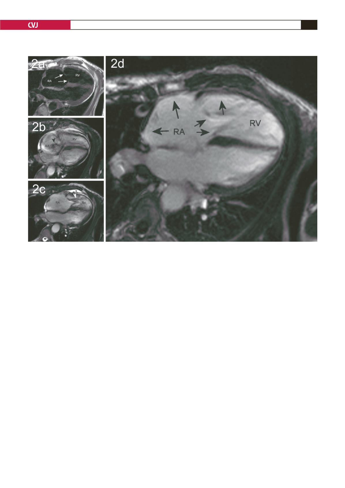
The right atrium was severely enlarged, while the leaflets of
the tricuspid valve were thickened and barely mobile (Fig. 2a).
The right ventricle was enlarged with diastolic flattening of the
interventricular septum due to right ventricle volume overload.
Signal voids caused by the turbulent flow through the tricuspid
valve during systole and diastole were observed, pointing to
Fig. 2. Magnetic resonance imaging. FSE T1-weighted black-blood four-chamber-view image presenting thickened and
retracted tricuspid valve leaflets (white arrows, a). Systolic (b), and diastolic (c) phase of ciné SSFP four-chamber-
view images demonstrating the signal void of tricuspid regurgitation (black arrowhead) and tricuspid stenosis (white
arrowhead). A small pericardial effusion (asterisk) and enlargement of the right atrium (RA) and right ventricle (RV)
were also observed. Endocardial delayed enhancement of the right atrium, tricuspid valve leaflets and right ventricle
(black arrows, d).


