CARDIOVASCULAR JOURNAL OF AFRICA • Vol 23, No 4, May 2012
AFRICA
e1
Case Report
Non-obstructive membranes of the left atrial appendage
TANIA BORDONALI, ALBERTO SAPORETTI, ENRICO VIZZARDI, ANTONIO D’ALOIA, ERMANNA CHIARI,
LIVIO DEI CAS
Abstract
The left atrial appendage (LAA) is a blind-ending, complex
structure distinct from the body of the left atrium and is
sometimes regarded as a minor extension of the atrium.
However, it should routinely be analysed as part of a trans-
oesophageal echocardiographic examination. In this study
we describe the presence of a non-obstructive membrane
traversing the cavity of the LAA, found incidentally on trans-
oesophageal echocardiography.
Keywords:
left atrial appendage, transoesophageal echocardiog-
raphy, left atrial thrombosis
Submitted 6/6/10, accepted 31/5/11
Cardiovasc J Afr
2012;
23
: e1–e2
DOI: 10.5830/CVJA-2011-020
A 70-year-old man without traditional risk factors presented
to our emergency room with palpitations and dyspnoea. A
12-lead electrocardiogram showed atrial fibrillation with a high
ventricular rate. Two-dimensional echocardiogram revealed a
hypertrophic left ventricle with reduced left ventricular function
(ejection fraction 45%). Pre-cardioversion transoesophageal
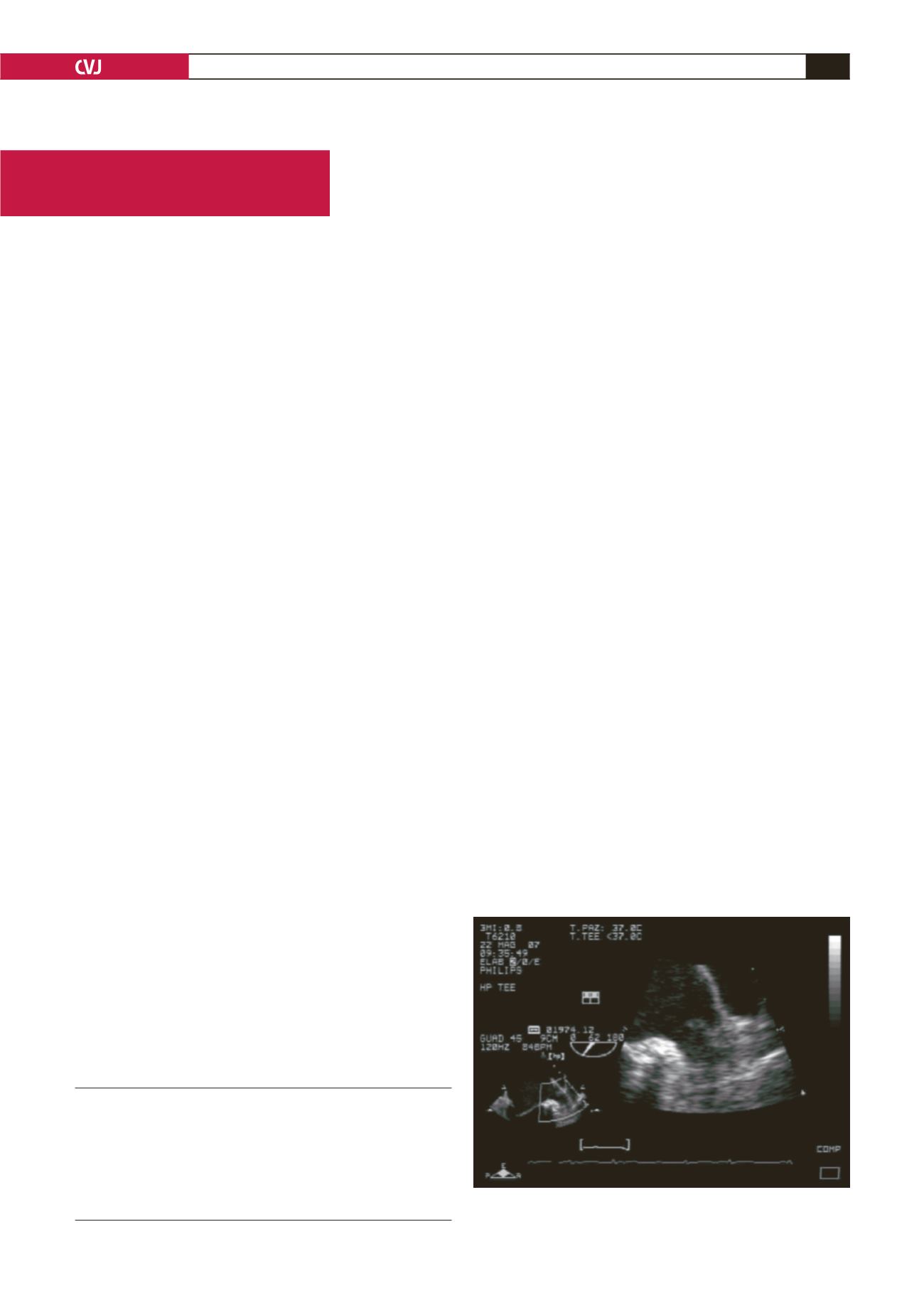
echocardiography (TEE) showed an enlarged left atrial appendage
(LAA) with moderate spontaneous echo contrast and a long, thin
structure traversing the body of the LAA without obstructing it.
This image was first ascribed to a thrombus, so oral
anticoagulation was started. After four weeks on good
anticoagulation, a second TEE was performed and the same
image with the same dimensions was noted. After electrical
cardioversion the patient recovered his ventricular function and
was discharged on standard therapy.
Discussion
When investigating the LAA by TEE, it is important to keep
in mind that the LAA is a three-dimensional (3D), multi-lobed
structure.
1-8
Therefore, evaluation should include imaging in
multiple planes, including orthogonal views, in order to image
the entire complex 3D structure.
Measurement of the two-dimensional (2D) LAA area is
not reproducible or helpful, in view of this complex structure.
Improved imaging techniques and the use of biplane and
multi-plane TEE have allowed visualisation of the LAA, which
previously was difficult to do using other imaging methods.
The presence of membranes on the left LAA cavity is rare, and
their origin is not clear.
9
Embryologically, the trabecular LAA is
a remnant derived from the left wall of the primary atrium,
which forms during the third and fourth weeks of embryonic
development.
10
The main smooth-walled left atrial cavity develops
later and is formed from an outgrowth of the pulmonary veins.
10
Reviewing variations in size and morphological characteristics
of the LAA,
1,3,7,8
no mention was made of these membranes.
When performing a TEE in patients with atrial fibrillation,
the pectinate muscles should not be confused with a thrombus.
Because the accuracy of LAA thrombus detection with TEE is
important in the pre-cardioversion evaluation of these patients, it
is vital to know what variations, especially in location and size
of the pectinate muscle, exist in the normal anatomy of the LAA.
Larger pectinate muscles (
≥
1 mm) occur in 97% of LAAs and
constitute another potential pitfall in TEE imaging of the LAA.
Small (
<
1 mm) pectinate muscles are seen only in the first and
last decades of life.
11
The differential diagnosis of long, thin structures in the LAA
may therefore include prominent pectinate muscles, side lobe
artefacts and partial resolution of thrombi. The images in our
case, however, seemed different.
The clinical origin and implications of these membranes are
not certain, however, they may represent an anatomical variant
that the echocardiographer should be aware of.
Istitute of Cardiology, University of Brescia, Spedali Civili
Brescia, Italy
TANIA BORDONALI, MD,
ALBERTO SAPORETTI, MD
ENRICO VIZZARDI, MD
ANTONIO D’ALOIA, MD
ERMANNA CHIARI, MD
LIVIO DEI CAS, MD
Fig. 1. Left atrial appendage in transoesophageal echo-
cardiography.


