
CARDIOVASCULAR JOURNAL OF AFRICA • Vol 24, No 2, March 2013
AFRICA
31
cases, 27.7%). Other lesions included MR + plus mitral stenosis
(MS) + AR (11 cases, 8.5%), MR + MS (nine cases, 6.9%), pure
MS (nine cases, 6.9%), MS + AR (seven cases, 5.4%), MR + MS
+ AR + aortic senosis (AS) (two cases, 1.5%) and MS + AS +
AR (one case, 0.8%).
The mitral valve was involved in all cases, while 73 cases
(56.2%) had isolated mitral valve lesions. All patients who
had aortic valve disease had associated mitral valve disease
(57 cases, 43.8%). Only one case was found to have abnormal
morphology (thickening) of the tricuspid valve. Isolated MR or
in association with AR was the most common finding detected
among children and adolescents (100% of age group
<
12 years
and 85% of 12–19 years). MS was less frequent in these age
groups. Although MS, AS and multiple valve lesions appeared
in adolescents, their frequency increased in young adult patients.
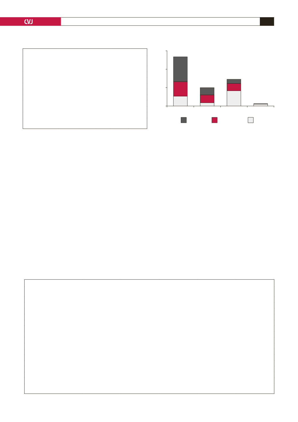
Fig. 4 shows the degree of severity of RHD according to
valvular lesion. The four types of valvular lesions were found in
mild, moderate and severe forms; 72.9% of the lesions fell into the
moderate and severe degree. Moreover, in mild-degree lesions,
AR prevailed, while in the severe form, MR was predominant.
Table 3 shows the echo features and complications of the study
population according to the predominant rheumatic valvular
lesion. Patients having haemodynamically significant valvular
disease affecting two valves were counted twice. Presentation
with an impaired systolic function of a left ventricular ejection
fraction
<
55% was not uncommon; 20 of 112 cases (17.9%)
presenting with mitral regurgitation and five of 28 cases (17.9%)
presenting with aortic regurgitation.
Chamber dilatations were much more frequent findings than
systolic dysfunction; left ventricular dilatation was seen in 60
(46.2%) cases, with a predominant proportion with MR (53.6%)
and AR (56.2%). Prevalence of left atrial dilatation in the study
group was as high as 75.4%. Pulmonary hypertension (53.3%)
was the most detected complication by echo and more related
to mitral lesions (both MR and MS). Other complications were
less frequent. Atrial fibrillation (13.9%) was the characteristic
complication of MS. Infective endocarditis was found in 10
(7.7%) cases, and was mainly associated with MR and AR.
Although no patient presented with a history of stroke or
left atrial thrombus in our study group, three of the patients had
echo findings of left atrial spontaneous echo contrast, which
carries a very high risk of left atrial thrombus formation and
cardiovascular accident (stroke). Almost half of the patients
presented with clinical heart failure in NYHA class III and IV.
This was more prevalent in patients with mitral valve lesions.
Eight (6.2%) patients had evidence of recurrent ARF and 93
cases (71.5%) required valvular surgery, according to the NHFA/
CSANZ 2006 guidelines of management of RHD.
9
TABLE 2. DISTRIBUTION OFVALVE LESION BYAGE GROUP
All
<
12
years
12–19
years
20–39
years
40–65
years
Valve lesion(s)
130 (100%)
7
20
72
31
MR
55 (42.31) 4 (57.14) 12 (60) 26 (36.11) 12 (38.71)
MS
9 (6.92)
0
0
7 (9.72)
2 (6.45)
MR + MS
9 (6.92)
0
1 (5)
6 (8.33)
2 (6.45)
MR + AR
36 (27.69) 3 (42.86) 5 (25) 20 (27.78) 8 (25.81)
MS + AR
7 (5.38)
0
0
4 (5.56)
3(9.68)
MR + MS + AR
11 (8.46)
0
1 (5)
6 (8.33) 4 (12.90)
MS + AS + AR
1 (0.78)
0
0
1 (1.39)
0
MR + MS + AR + AS 2 (1.54)
0
0
2 (2.78)
0
MR
=
mitral regurgitation; MS
=
mitral stenosis; AR
=
aortic regurgitation; AS
=
aortic stenosis.
TABLE 3. ECHOCARDIOGRAPHIC FEATURESAND COMPLICATIONSACCORDINGTO PREDOMINANTVALVULAR LESION
Total cases
Total
130 (100%)
MS
30 (23.08%)
MR
112 (86.15%)
AS
1 (0.77%)
AR
28 (21.15%)
Echo features
Mean LVEF
61.15
63.96
59.85
67
48.76
Systolic dysfunction
27 (20.77)
2 (6.67)
20 (17.86)
0
5 (17.86)
Mean LVIDD
55.89
43.9
57.57
55
62.69
LV dilatation
60 (46.15)
3 (10)
60 (53.57)
0
15 (53.57)
Mean LA
50.48
52.02
49.35
50
50.46
LA dilatation
98 (75.38)
25 (83.33)
78 (69.64)
1 (100)
24 (85.71)
Spontaneous echo contrast
3 (2.31)
1 (3.33)
2 (1.79)
0
2 (7.14)
Complications
PHT
73 (56.15)
16 (53.33)
56 (50)
0
13 (46.43)
IE
10 (7.69)
1 (3.33)
8 (7.14)
0
3 (10.71)
AF
18 (13.85)
7 (23.33)
11 (9.82)
0
2 (7.14)
NYHA class III/IV
56 (43.08)
13 (43.33)
42 (37.5)
0
9 (32.14)
Definite recurrent ARF
4 (3.08)
1 (3.33)
3 (2.68)
0
0
Probable recurrent ARF
4 (3.08)
0
4 (3.57)
0
0
LV = left ventricle; LVEF = left ventricular ejection fraction; LVIDD = left ventricular internal diameter in diastole; LA = left atrium; PHT = pulmonary hyperten-
sion; IE = infective endocarditis; AF = atrial fibrillation; NYHA = NewYork Heart Association; ARF = acute rheumatic fever.
Fig. 4. Severity of valve lesions.
120
100
80
60
40
20
0
Severity of lesions
MR
MS
AR
AS
severe
moderate
mild
Valvular lesions


