CARDIOVASCULAR JOURNAL OF AFRICA • Vol 24, No 6, July 2013
e2
AFRICA
vascular graft (Hemashield, Boston Scientific, Boston, MA,
USA) with new vascular ring connectors (VRC). The VRC
was attached to the vascular prosthesis by securing the edge
of the graft against the narrow groove of the VRC with
non-absorbable sutures. The ringed prosthesis was then inserted
into the abdominal aorta, positioned at the planed proximal
anastomotic site, and fixed to the abdominal aorta by tying the
tapes against the wider groove. A similar manoeuvre was used
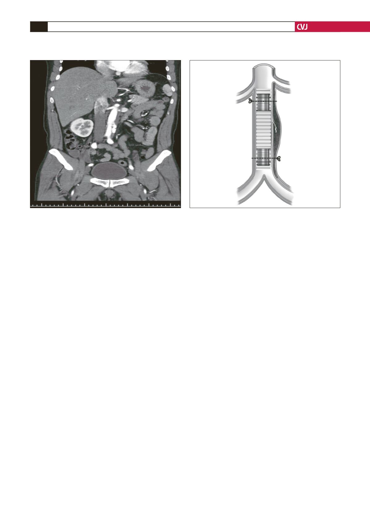
for the distal anastomosis. The patient was well without deficit
at the one-year follow up (Fig. 2).
Discussion
IMH results either from spontaneous rupture of the vasa
vasorum of the aorta, causing haemorrhage within the aortic
wall in the absence of initial intimal disruption, or from a
penetrating atherosclerotic ulcer that penetrates into the internal
elastic lamina and allows haematoma formation within the
media of the aortic wall.
4-7
IMH is a variant form of aortic
dissection, and although less common, accounts for 10–30%
of cases. It has been accepted as an increasingly recognised
and potentially fatal entity of all acute aortic syndromes.
4,7
Penetrating atherosclerotic ulcer of the IMH was reported to
have a significantly progressive course.
5,6
In our experience,
IMH due to a penetrating atherosclerotic ulcer commonly had a
progressively downhill clinical course.
IMH has a variable clinical course. It commonly affects elderly
patients (mean age 66 years) with a history of hypertension.
5,6,8
The most common presenting symptom is sudden onset of
abdominal pain radiating to the back or the buttock.
5
In our
experience, onset of recurrent/refractory pain may be a sign of
impending rupture and should therefore be considered for more
aggressive intervention.
With recent advances in imaging techniques, IMH is now
increasingly recognised. CT scans diagnose aortic dissection
reasonably well but may not be completely reliable in
distinguishing IMH from classic aortic dissection.
8
Aortography
was performed, which showed no evidence of dissection but
did reveal an ectatic descending aorta. With MRI, IMH is
characteristically seen as a focal thickening of the aortic wall
in the absence of dissection.
8
It should be considered in the
differential diagnosis of any patient with an acute onset of
abdominal pain, radiation to the back or buttocks, with the
presence or absence of a pulsatile abdominal mass or signs of
limb ischaemia.
Most authorities currently recommend treatment of IMH
similar to that of classic aortic dissection, with early surgery
for patients with proximal IMH and medical management for
patients with distal IMH.
8
Initially stable patients with IMH
of the abdominal aorta can be treated medically with frequent
clinical and radiological re-assessment and they may have
complete resolution.
A certain volume of haematoma in the aortic wall may cause
the intima of the penetrating atherosclerotic ulcer to become
fragile and lead to intimal disruption.
1
Consequently IMH
weakens the aorta and may progress to aortic dissection. This
may explain why wall thickness is related to progression to
overt dissection or aortic rupture. IMH may progress to classic
dissection (i.e. with intimal disruption) in up to 33% of cases.
5,8
In our case, intramural haematoma of the abdominal aorta
progressed to rupture with medical failure.
Conservative medical management is usually the first choice
of treatment for Stanford type B acute aortic dissection without
complications, but surgical intervention is required in patients
with complications, such as leakage or threatened rupture,
malperfusion, persistent pain or intractable and progressive
enlargement of the ulcer-like projection to more than 20 mm in
diameter or depth during the follow-up period.
1,2,8
Operative intervention includes open or endovascular repair
of the abdominal aorta. The open technique usually involves
Fig. 1. A follow-up contrast-enhanced CT showing aortic
dissection with a large primary entry tear in the infra-
renal site of the abdominal aorta, extending to the bifur-
cation of the distal abdominal aorta on day 14 after onset.
Fig. 2. The ringed graft is inserted into the aorta and the
braided nylon tapes are tied against the wider groove
of the vascular ring connectors to achieve a sutureless
anastomosis.


