


 25 / 64

CARDIOVASCULAR JOURNAL OF AFRICA • Volume 30, No 4, July/August 2019
AFRICA
211
blood vessels stain blue-black and red/pink, respectively. Sections
with more pink and black stain represent increased collagen and
elastic fibres, as shown in Fig. 6.
Sections from the arch and abdominal aorta showed more
deposition of collagen and elastic fibres in the intima of pups
born to hypercholesterolaemic mothers in the HC group, but
Treatment groups of rabbits
NC
HC
HCC
Intima–media thickness (mm)
0.2
0.1
0.0
*
*#
Fig. 3.
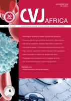
(A) shows micrographs of H & E-stained sections of the aortic arch of rabbit pups born to the control rabbits (NC), hyper-
cholesterolaemic rabbits without cocoa (HC) and hypercholesterolaemic rabbits given cocoa (HCC). The value of the scale
bar is 140 µm. (B) shows a bar chart of the intima–media thickness of the aortic arch of offspring from the three groups. *
p
<
0.001 and
#
p
<
0.001 compared to the HC group. Error bars indicate standard deviation.
A
B
NC
HC
HCC
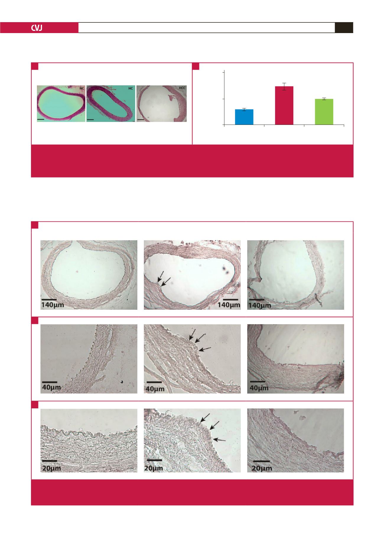
Fig. 4.
Micrographs at different magnifications of Oil red O-stained sections of the aortic arch of offspring. (A) shows a whole section
of the aortic arch. (B) and (C) show higher magnifications of the aortic arch and lesions (indicated with arrows). Sections from
the NC and HCC pups show no lesions, whereas sections from the HC pups show the presence of atherosclerotic lesions.
A
B
C