CARDIOVASCULAR JOURNAL OF AFRICA • Vol 22, No 2, March/April 2011
AFRICA
91
was first described as an entity in 1990 by Chin
et al
.
1,2
It
occurs because of an arrest in the normal embryonic process of
myocardial compaction, which occurs after the fourth week of
life. This results in the persistence of trabeculae together with
adjacent deep intra-trabecular recesses filled with blood. The
term ‘isolated’ is reserved for situations in which there are no
associated cyanotic congenital heart disease, obstructive intra-
cardiac lesions, anatomical valvular abnormalities or associated
coronary anomalies. This process may involve the right ventricle
in less than 50% of cases.
3
The diagnosis on echocardiography is suspected by the pres-
ence of more than three trabeculations, usually in the apex,
apicolateral and apico-inferior walls. The apical and mid-ventric-
ular walls of the inferior and lateral walls can be involved in up
to 80% of cases. Deep intra-trabecular recesses accompany this,
and the presence of blood flow within these recesses on colour
Doppler is essential. The Jenni criteria utilise these features as
the basis for diagnosis on two-dimensional echo, emphasising
that on parasternal short axis, the ratio of the non-compacted to
compacted layer should be greater than 2 at end-systole.
4,5
Recently, a few case reports suggested that three-dimensional
echo may improve diagnosis in subtle cases.
6
Contrast echo is
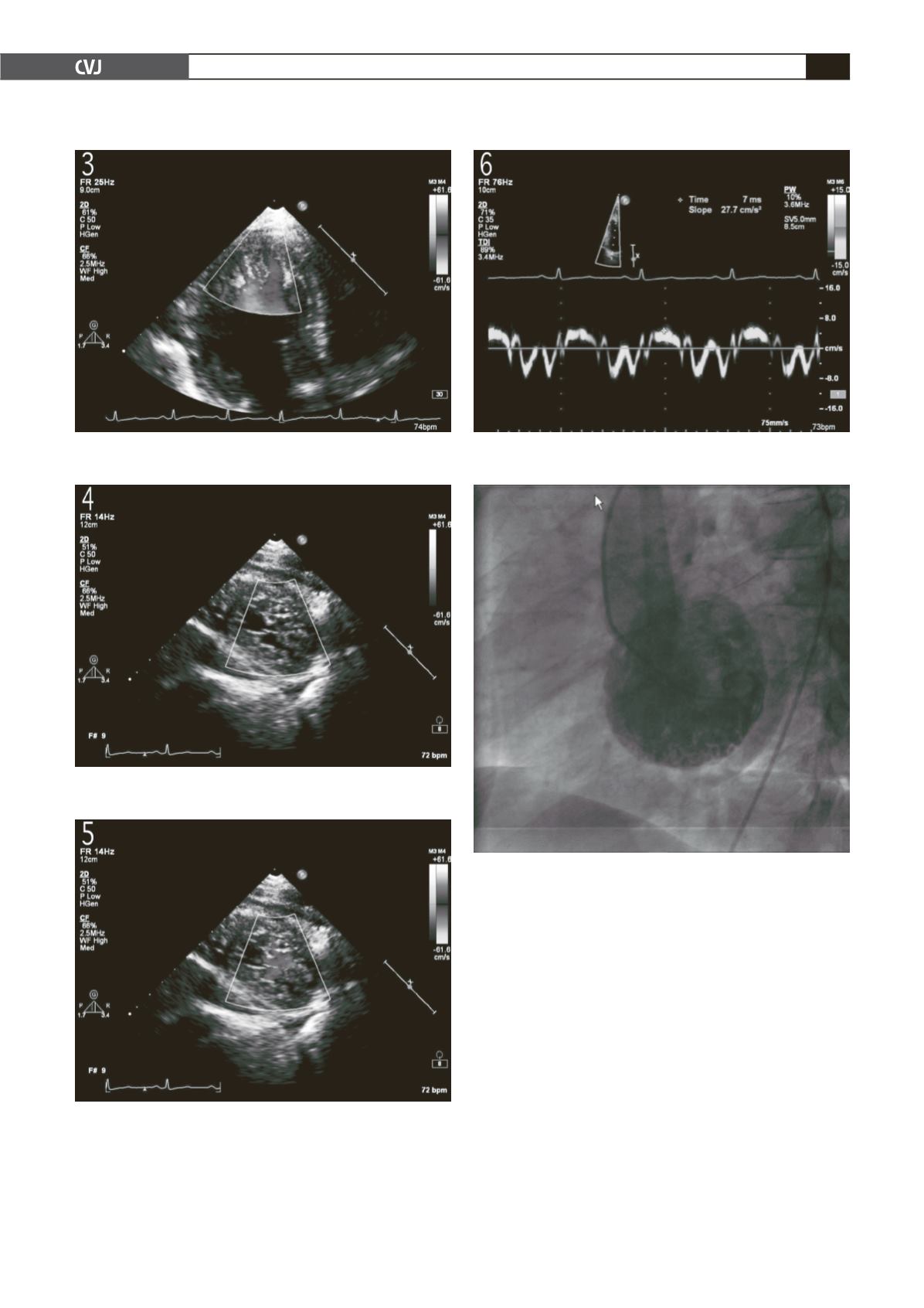
Fig. 3. Apical two-chamber in diastole demonstrating flow
within the trabeculae.
Fig. 4. Short-axis view at the apex demonstrating marked
trabeculation.
Fig. 5. Short-axis view at the level of the apex demon-
strating flow within the trabeculae.
Fig. 6. Tissue Doppler demonstrating a reduced E
′
in
keeping with diastolic dysfunction.
Fig. 7. Left ventriculargram demonstrating marked
trabeculation with dye staining in apex and lateral wall.


