CARDIOVASCULAR JOURNAL OF AFRICA • Vol 22, No 2, March/April 2011
94
AFRICA
carboxyhaemoglobin level was 22%.
The patient was intubated and placed on 100% oxygen.
Five hours after intubation, she was stabilised and extubated.
On extubation, the patient had no angina pectoris or dyspnoea.
Hyperbaric oxygen treatment was applied at 2.5 atm pressure for
150 minutes. The carboxyhaemoglobin level reduced to 2.5% by
the twelfth hour post admission.
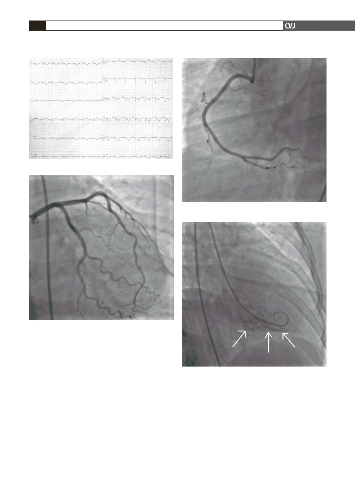
Although the patient did not have any symptoms, an ECG was
done and it revealed negative T waves in leads D2, D3 and avF,
ST-segment elevation on avR, 1 mm ST-segment depression and
symmetric T wave negativity in leads V3–6, D1 and avL (Fig.
1). Serum cardiac markers were re-evaluated, and creatine kinase
(CK) was 220 U/l, CK-MB 32 U/l and troponin I 2.6 ng/ml. The
infero-apical segment was hypokinetic on echocardiography.
Coronary angiography was performed on the same day and
revealed normal coronary arteries (Figs 2, 3), however the infero-
apical region was also hypokinetic on left ventriculography (Fig.
4). The patient remained asymptomatic on anti-thrombotic and
Fig. 1. Ischaemic aberrations in several leads on the ECG.
Fig. 2. Right anterior oblique coronary angiographic view
with caudal angulation demonstrating normal left ante-
rior descending and circumflex coronary arteries.
vasodilatator treatment. The ischaemic aberrations on the ECG
normalised on the sixth day, without pathological Q waves in the
inferior derivations (Fig. 5).
The patient was discharged on the sixth day without compli-
cations and a Tc-99m SPECT performed one month later did not
reveal myocardial necrosis or ischaemia. A control echocardio-
graphy revealed normalisation of the abnormality in the myocar-
dial segmentary wall motion.
Discussion
When the relationship between myocardial oxygen supply
and demand is disturbed, cardiac ischaemia occurs. Although
Fig. 4. Right oblique projection of left ventriculogram
revealing infero-apical hypokinesia.
Fig. 3. Left anterior oblique coronary angiographic view
showing normal right coronary artery.


