CARDIOVASCULAR JOURNAL OF AFRICA • Vol 22, No 2, March/April 2011
AFRICA
95
coronary artery disease is the most common reason for cardiac
ischaemia, this does not explain the nature of the ischaemic
event in many clinical situations.
1
Increased viscosity and altered
platelet function have been proposed as the pathophysiological
mechanisms in patients with acute myocardial infarction with
normal coronary arteries.
The affinity of CO to haemoglobin is 200 to 270 times greater
than that of oxygen; therefore, the formation of carboxyhaemo-
globin not only decreases the amount of oxygen delivered to the
tissues but also displaces the oxygen–haemoglobin dissociation
curve to the left. Cardiac toxicity may result from myocardial
hypoxia or from the direct toxic effect of CO on the myocardial
mitochondria. An increased tendency for thrombosis and coro-
nary vasospasm are also responsible for myocardial damage in
patients with CO poisoning.
2
Myocardial infarction has been
reported in patients with underlying CAD.
3
Cardiac involve-
ment may occur promptly after exposure, or may be delayed for
several days, such as in our presented case.
Electrocardiographic abnormalities have been described with
acute carbon monoxide poisoning in human and animal models.
These include premature atrial and ventricular contractions,
4-6
infranodal
7
and intraventricular blocks
8
and anoxic disorders in
the ST segment and T wave.
9
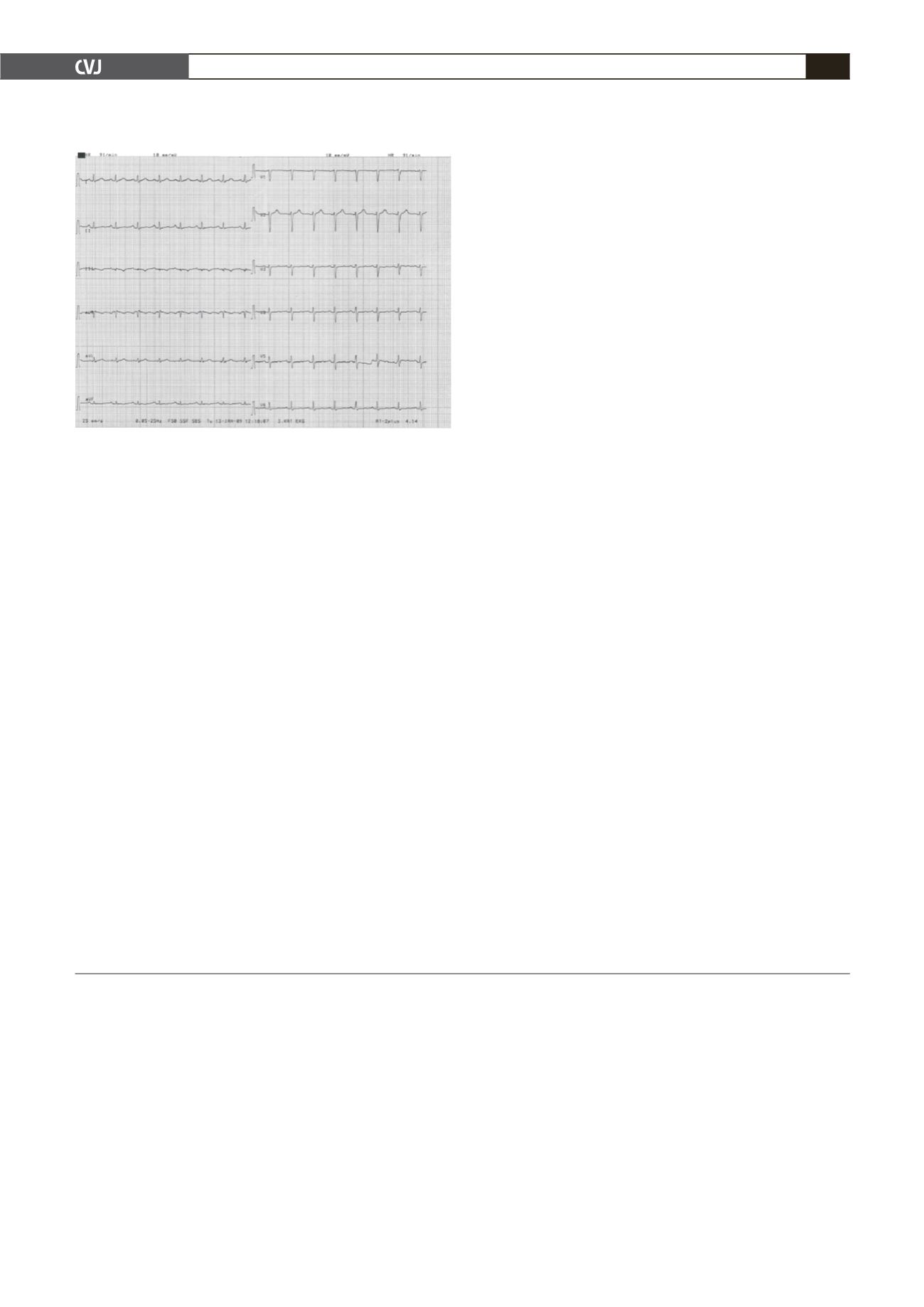
Although in our patient, angio-
graphically normal coronary arteries, infero-apical hypokinesia
and troponin I levels suggested prolonged vasospasm or spon-
taneous lysis of an intracoronary thrombus as the responsible
mechanism of the reversible myocardial stunning, diffuse precor-
dial T wave negativity and ST segment elevation of avR denoted
diffuse myocardial ischaemia as a concomitant mechanism.
Conclusion
Frequent ECG and cardiac enzyme monitoring is important in
the management of patients with CO poisoning, as asymptomatic
myocardial ischaemia and reversible myocardial stunning may be
observed in the acute phase or several days later.
References
1. Rosenblatt A, Selzar A. The nature and clinical features of myocar-
dial infarction with normal coronary arteriogram.
Circulation
1977;
55
:
578–580.
2. Marius-Nunez AL. Myocardial infarction with normal coronary arteries
after acute exposure to carbon monoxide.
Chest
1990;
97
: 491–494.
3. Varol E, Ozaydin M, Aslan SM, Do
ğ
an A, Altinba
ş
A. A rare cause
of myocardial infarction: acute carbon monoxide poisoning.
Anadolu
Kardiyol Derg
2007;
7
(3): 322–323.
4. Shafer N, Smilay MG, MacMillan FR. Primary myocardial disease man
resulting from acute carbon monoxide poisoning.
Am J Med
1965;
38
:
316–320.
5. Stearns WH, Drinker CK, Shaughnessy TJ. The electrocardiographic
changes found in 22 cases of carbon monoxide poisoning.
Am Heart J
1938;
14
: 434–446.
6. Aslan S, Erol MK, Karcıo
ğ
lu O, Meral M, Çakır Z, Katırcı Y. The
investigation of ischemic myocardial damage in patients with carbon
monoxide poisoning.
Anadolu Kardiyol Derg
2005;
5
: 189–193.
7. Ehricb WE, Bellet S, Lewey Fli. Cardiac changes from carbon monoxide
poisoning.
Am J Med Sci
1944;
208
: 511–521.
8. Colvin LT. Electrocardiographic changes in cases of severe carbon
monoxide poisoning.
Am Heart J
1928;
3
: 484.
9. Middleton GD, Ashby DW, Clark F. Delayed and long-lasting electro-
cardiographic changes in carbon monoxide poisoning.
Lancet
1961;
1
:
12–14.
Fig. 5. Normalised ECG findings.


