CARDIOVASCULAR JOURNAL OF AFRICA • Vol 22, No 2, March/April 2011
90
AFRICA
Case Reports
Isolated left ventricular non-compaction with normal
ejection fraction
F PETERS, C DOS SANTOS, R ESSOP
Summary
Isolated left ventricular non-compaction (LVNC) is a genetic
disease that is being increasingly recognised in patients
presenting with heart failure of unknown origin. In this case
report, we describe a patient with classic LVNC without
clinical heart failure and with normal left ventricular ejec-
tion fraction.
Keywords:
normal systolic function, isolated left ventricular
non-compaction
Submitted 18/11/09, accepted 14/3/10
Cardiovasc J Afr
2010;
21
: 90–93
DOI: CVJ-21.017
A 37-year-old lady presented with atypical chest pain and was
referred by her primary physician for an echocardiogram to
evaluate her cardiac structure and function. On direct question-
ing, she had no symptoms to suggest cardiac dysfunction in
particular, nor did she have any symptoms suggestive of an
arrhythmia. Her physical examination was unremarkable except
for tenderness over her left costal cartilages, in keeping with
costochondritis. The resting electrocardiogram was normal.
An echocardiogram revealed that she had normal cardiac
dimensions measured at the base of the heart, with a global ejec-
tion fraction of 60%. There were no regional wall abnormalities
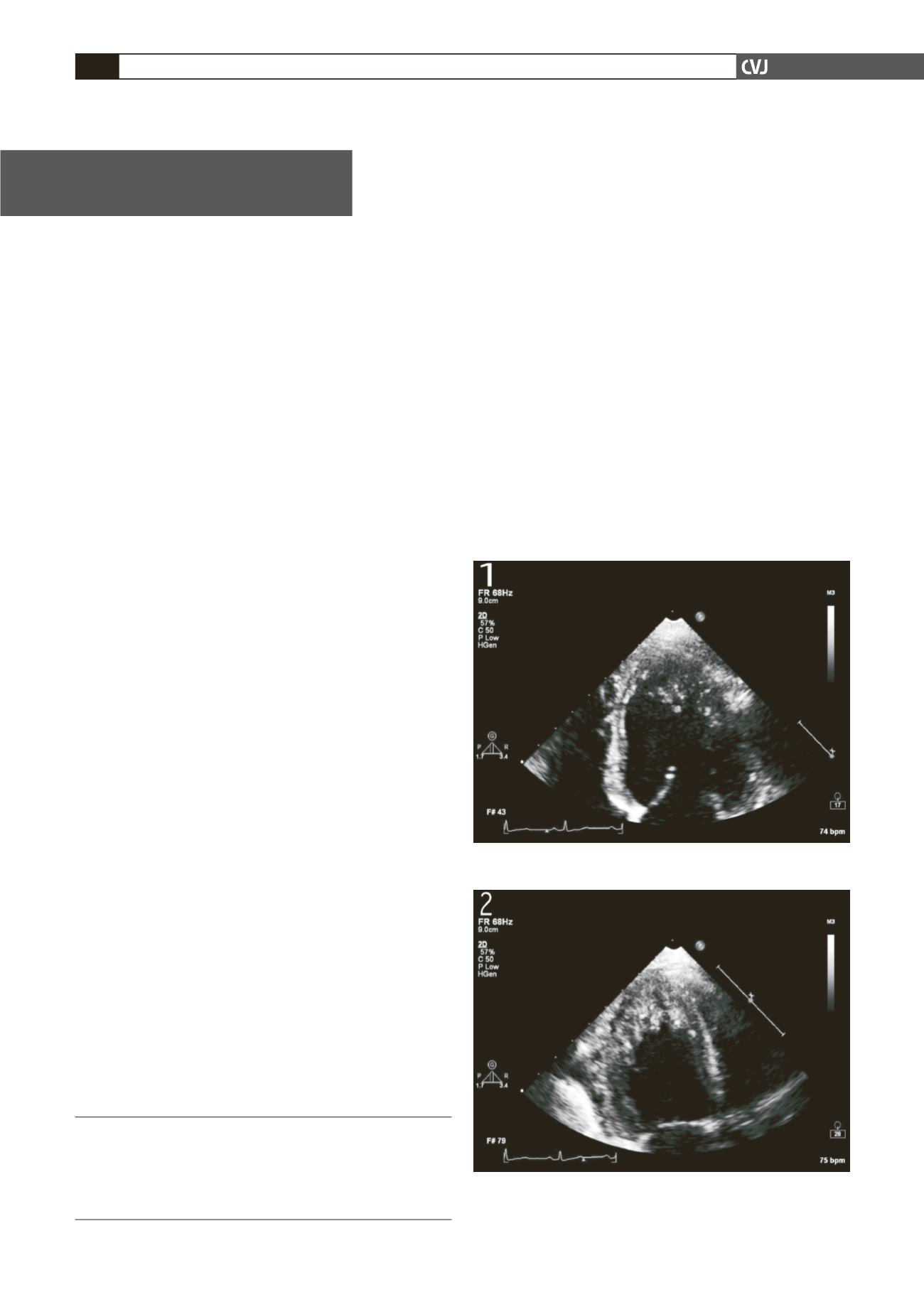
but she had marked trabeculation involving the apex, apicolateral
and apico-inferior walls (Figs 1, 2, 4). Deep intra-trabecular
recesses with flow were demonstrated using colour Doppler
(Figs 3, 5). On the Jenni criteria, the ratio of non-compacted to
compacted myocardium was greater than 2, measured at end-
systole in the parasternal short-axis view. Mitral inflow Doppler
revealed a pseudo-normalisation pattern with a diminished E
′
on
tissue Doppler, suggesting grade 2 diastolic dysfunction (Fig. 6).
The right ventricle was normal. The rest of the heart was normal
structurally and functionally.
Coronary angiography revealed no anomalies, with normal
epicardial coronary anatomy and flow. The left ventricular
angiogram revealed marked trabeculation with dye staining in
the same areas noted on the echocardiogram (Fig. 7). The ejec-
tion fraction was 65%. A Holter electrocardiogram was normal.
A final diagnosis of isolated left ventricular non-compaction
with preserved left ventricular systolic function and moder-
ate diastolic dysfunction was made. The patient was placed on
warfarin.
Discussion
Isolated LV non-compaction of the myocardium
Isolated LV non-compaction was classified as a genetic cardio-
myopathy in the 2006 classification of cardiomyopathy, and
Division of Cardiology, Chris Hani Baragwanath Hospital,
Johannesburg, South Africa
F PETERS, MB BCh, FCP (SA), Cert Cardiol (SA),
C DOS SANTOS, BSc
R ESSOP, MB BCh, FCP (SA), FACC, FRCP (London)
Fig. 1. Apical four-chamber view in diastole demonstrat-
ing trabeculation of the apex and apicolateral wall.
Fig. 2. Apical two-chamber view in end-systole demonstrat-
ing the marked trabeculation of the apex and apico-inferior
wall with the underlying thin epicardial compacted area.


