CARDIOVASCULAR JOURNAL OF AFRICA • Vol 23, No 9, October 2012
AFRICA
487
p
Ds
<
0.001),
mean velocity (
p
Ab
<
0.001,
p
Ds
<
0.001),
mean PG
(
p
Ds
<
0.001),
D/S ratio velocity (
p
Ab
<
0.001,
p
Ds
<
0.001),
VTI
(
p
Ab
=
0.005,
p
Ds
<
0.001)
and time to peak systolic velocity (
p
Ab
<
0.001,
p
Ds
=
0.026).
While the PSV of the abdominal aorta
was significantly increased after stenting (
p
Ab
<
0.001),
the
corresponding value of the descending aorta had decreased (
p
Ds
<
0.001).
The largest percentage increase (119.57%) was achieved
in the pulsatility index, which increased from 0.89
±
0.30
to 1.75
±
0.51 (
p
Ds
<
0.001).
The ROC curve analysis was performed to evaluate the
diagnostic values of different Doppler echocardiographic indices
in order to differentiate significant coarctation (pre-stenting
condition) from post-stenting conditions or without coarctation.
Diagnostic values of all 12 Doppler echocardiographic indices
in both the abdominal and descending aorta are given in Table 3.
As shown in Table 3, except for the mean velocity (
p
Ab
=
0.177)
and mean PG (
p
Ab
=
0.489)
of the abdominal aorta, other
indices of the abdominal or descending aorta had a statistically
significant area under the curve (AUC) to distinguish patients
with significant aortic coarctation or a pre-stenting condition.
The VTI of the descending aorta had the greatest AUC of 0.980
(
p
Ds
<
0.001)
such that the velocity–time integral of
>
87.50
had
90.9%
sensitivity and 91.3% specificity, and the values of
>
94.50
had 86.4% sensitivity and 100% specificity for detection
of significant aortic coarctation.
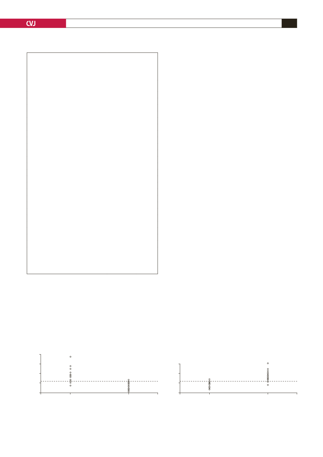
A pulse delay of
>
5.98
had a sensitivity of 87% and
specificity of 95.7% to diagnose significant aortic coarctation
(
Fig. 3). Moreover, as illustrated in Fig. 4, a pulsatility index of
>
1.21
had 87% sensitivity and 91.3% specificity to differentiate
significant coarctation (pre-stenting condition) from the post-
stenting condition or without coarctation.
As shown in Table 4, the baseline peak aortic gradient
measured by catheter was significantly correlated with some
of the pre-stenting echocardiographic profiles of the abdominal
and descending aorta. In both the abdominal and descending
aorta, the strongest correlation of the peak gradient was observed
with pressure half-time as a direct relationship (
r
Ab
=
0.637,
p
Ab
=
0.001;
r
Ds
=
0.613,
p
Ds
=
0.003).
The velocity–time integral
of the descending aorta was also directly correlated with
before-stenting peak gradients (
r
Ds
=
0.548,
p
Ds
=
0.010).
Other
correlations are shown in Table 4.
The possible correlation of the baseline peak aortic gradient
with the mean percentage change in echocardiographic profiles
after stenting was also evaluated. Our findings showed a reverse
correlation between severity of coarctation and changes in LDV
after stenting. The higher the gradient, the lower the change in
the LDV of the abdominal aorta (
r
Ab
=
–0.455,
p
Ab
=
0.033).
By
contrast, changes in the PHT of the abdominal aorta were directly
correlated with the baseline catheter gradient (
r
Ab
=
0.436,
p
Ab
=
0.043).
Changes in the other echocardiographic indices were not
significantly correlated with the baseline aortic gradient.
Discussion
Coarctation of the aorta is characterised by anatomical obstruction
in the descending aorta. It is difficult to evaluate this obstruction
because of the variability in cardiac output, number and size of
collaterals, and peripheral resistance.
25
Primary clinical diagnosis
and subsequent assessment of the severity of coarctation and
Fig. 3. Cut-off points of 5.98 for pulse delay of descend-
ing aorta to differentiate significant coarctation (pre-
stenting condition) from post-stenting condition or with-
out coarctation.
20
15
10
5
0
Pulse delay
Severe coarctation
(
pre-stenting condition)
Post-stenting
Fig. 4. Cut-off points of 1.21 for pulsatility index of
descending aorta to differentiate significant coarctation
(
pre-stenting condition) from post-stenting condition or
without coarctation.
3
2
1
0
Pulsatility index
Severe coarctation
(
pre-stenting condition)
Post-stenting
TABLE 4. EVALUATION OF THE CORRELATION BETWEEN
SEVERITY OF COARCTATION (BASELINE PEAK
GRADIENT OF THE CATHETER) AND PRE-STENTING
ECHOCARDIOGRAPHIC PROFILES OFABDOMINAL
AND DESCENDINGAORTA
Echocardiographic index
Correlation
coefficient (r) p-value
Abdominal aorta
PSV (m/s)
–0.093
0.681
EDV (m/s)
0.087
0.701
LDV (m/s)
–0.026
0.909
AT (m/s)
0.014
0.952
P.H.T (m/s)
0.637
0.001
Mean velocity (m/s)
0.066
0.769
Mean PG
0.115
0.610
D/S ratio velocity
0.054
0.811
VTI
0.446
0.038
Time to peak systolic velocity (m/s)
–0.004
0.987
Descending aorta
PSV (m/s)
0.265
0.245
EDV (m/s)
0.474
0.030
LDV (m/s)
0.592
0.005
AT (m/s)
0.093
0.688
PHT (m/s)
0.613
0.003
Mean velocity (m/s)
0.473
0.030
Mean PG
0.436
0.048
D/S ratio velocity
0.374
0.095
VTI
0.548
0.010
Time to peak systolic velocity (m/s)
–0.020
0.931
Pulse delay
–0.075
0.739
Pulsatility index
0.084
0.710
PSV: peak systolic velocity, EDV: early diastolic velocity, LDV: late
diastolic velocity, AT: systolic acceleration time, PHT: pressure half-time,
PG: peak gradient, D/S: diastolic velocity/systolic velocity, VTI: velocity
time integral, AUC: area under curve.
All data derived from two-sided Pearson correlation analysis.


