CARDIOVASCULAR JOURNAL OF AFRICA • Volume 25, No 1, January/February 2014
18
AFRICA
Discussion
There were three major findings of this study. The intra- and
inter-atrial, and intra-left atrial electromechanical coupling
intervals were significantly higher in the pregnant subjects.
The P
max
and PD were also significantly higher in the pregnant
subjects and there was a significant correlation between the
inter-atrial and the intra-left atrial electromechanical coupling
interval P
max
.
Clapp
et al
. reported a progressive increase in all cardiac
chamber dimensions in pregnancy.
,19
However, Katz
et al
.
found no statistically significant differences between left atrial
diameter and left ventricular internal diastolic diameter (LVIDD)
in pregnant subjects in the second trimester and at 12 weeks
postpartum.
,20
In our study, there were no differences between the
LA diameter and LVIDD in pregnant subjects compared to the
controls, which is in line with the previous study.
Increases in maternal blood volume, cardiac output and heart
rate are seen during pregnancy. These mechanisms affect the
refractory period and conduction velocity. Schwartz and Priori
found that stress and anxiety also caused arrhythmogenic effects
by acting on the sympathetic nervous system.
,21
Hormonal changes also seem to play an important role
in arrhythmias during pregnancy. Gleicher
et al
. found that
oestrogens increased the excitability and frequency of action
potentials in uterine muscle tissue during pregnancy.
,22
This
increased adrenergic sensitivity may play a role in the genesis of
arrhythmias by modifying the refractory period and conduction
velocity in the re-entrant circuit.
When left ventricular diastolic dysfunction occurs, emptying
of the left atrium is also impaired. Following impaired left
ventricular diastolic relaxation, there is increased atrial
contribution to the mitral flow in the left ventricular diastolic flow,
thus leading to myocardial overstretching and enlargement.
,23
In
our study, DT and A velocity were significantly higher in the
pregnant subjects, but IVRT, IRT and ET were similar to the
controls. The left atrium diameter is known to be correlated with
cardiovascular events and a risk factor for AF.
,24
These volumetric
changes also constitute a high risk for AF.
There are conflicting results on this topic. In our study, the
diameters of the pregnant subjects were similar to those of the
controls, in line with our previous studies,
24-26
whereas some
studies found increased LA dimension in patients with diastolic
dysfunction and increased coupling parameters.
27,28
We believe
there is a need for large-scale studies to shed light on this
discrepancy.
PD is related to non-homogenous and interrupted conduction
of sinus impulses intra- and inter-atrially. Currently, prolonged
P
max
, increased PD and atrial conduction disorders are associated
with a higher risk of paroxysmal atrial tachyarrhythmias.
7,8
Therefore, it has been suggested that PD can be used in the
diagnosis of patients with a high risk of AF.
7,8
It was moreover
shown that PD was prolonged in chronic, inflammatory and
rheumatic diseases, such as rheumatoid arthritis and Behcet’s
disease.
29,30
In another study, PD was increased in pregnancy due to
shortening of the minimum P-wave length and it reached its
longest length in the third trimester. Pregnancy also had no
effect on P
max
.
31
In our study, P
max
and PD were longer in pregnant
subjects than in the controls, and this increased PD was related
to prolonged P
max
.
There are several ways to measure total atrial conduction
time; one is signal-averaged ECG, which is the gold-standard
technique, but it requires special hardware and is a longer
technique. For this reason, measurement of total atrial conduction
time by signal-averaged ECG is not often used in clinical
practice.
Mercadier
et al.
have shown PA TDI to be an easy, fast
and reliable method to measure total atrial electrical activation
time.
,23
PA TDI duration is a readily available echocardiographic
tool to estimate total atrial conduction time and it can easily be
measured by all cardiologists. This novel echocardiographic tool
has been validated by the P-wave duration on signal-averaged
electrocardiography.
32
Atrial conduction time can be measured by both invasive and
non-invasive methods.
33
Prolongation of atrial conduction time,
as measured by TDI, is an independent predictor of new-onset or
recurrent AF.
34,3
Several studies have found that atrial conduction
time measured by TDI increases in patients with various
diseases, such as type 1DM,
25
dilated cardiomyopathy,
36
and
ankylosing spondilitis.
26
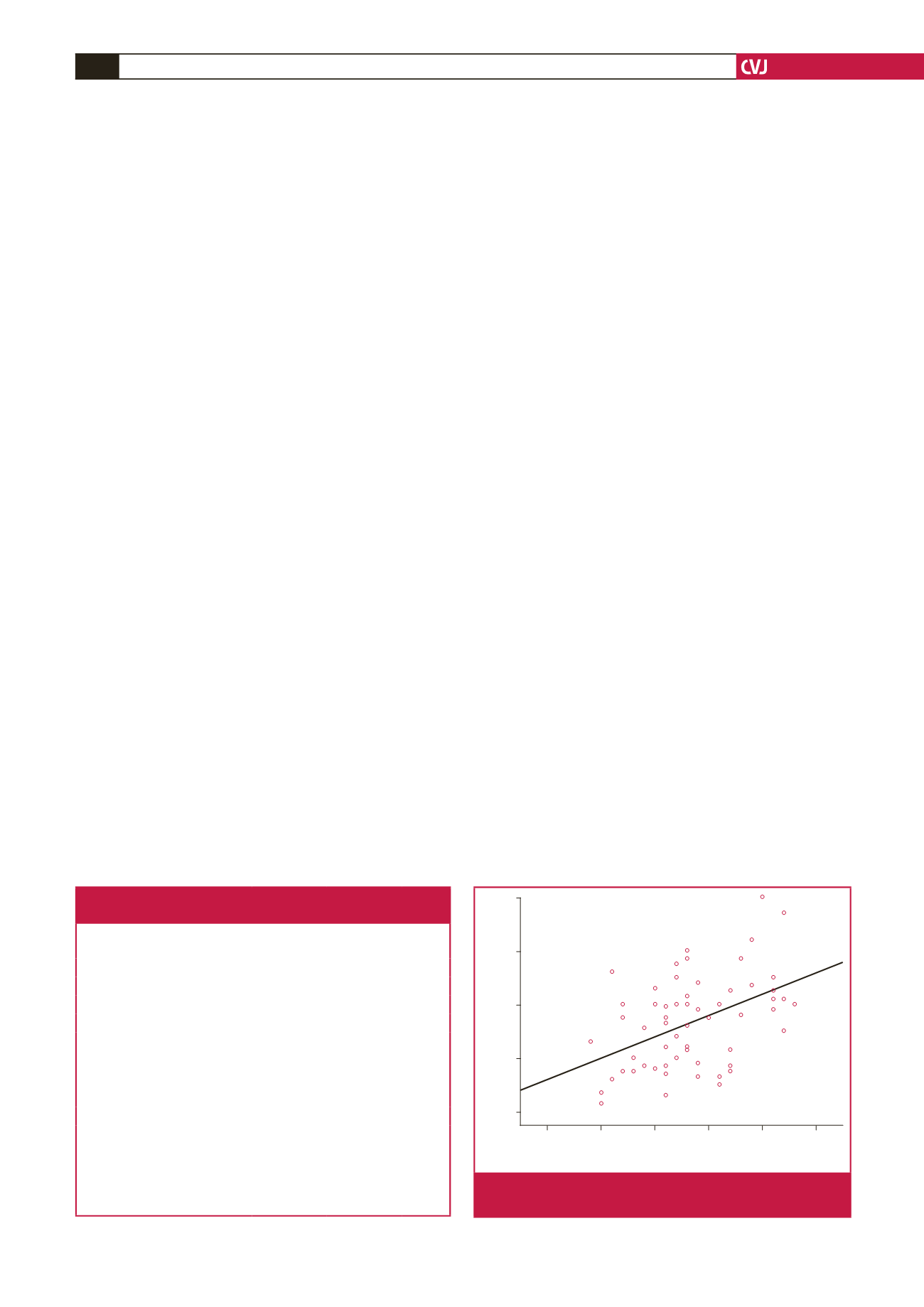
120
100
80
60
40
10.00 15.00 20.00 25.00 30.00 35.00
Inter-atrial electromechnical interval (ms)
A velocity (ms)
r
=
0.459
R
2
linear
=
0.21
Fig. 2.
Positive correlation between inter-atrial electro-
mechanical intervals and mitral A-wave velocity.
Table 3. Comparison of the electrocardiographic and
electromechanical coupling parameters
Pregnant
subjects
Controls
p
-value
Maximum P-wave duration (ms) 103.1
±
5.4 96.8
±
7.4
<
0.001
Minimum P-wave duration (ms)
52.4
±
6.3 55.1
±
5.7 0.090
P-wave dispersion (ms)
50.7
±
6.8 41.6
±
5.5
<
0.001
PA lateral (ms)
62.1
±
2.7 55.3
±
3.2
<
0.001
PA septal (ms)
45.7
±
2.5 43.1
±
2.7
<
0.001
PA tricuspid (ms)
35.7
±
2.7 35.1
±
3.2 0.440
PA lateral – PA tricuspid*
26.4
±
4.0 20.2
±
3.6
<
0.001
PA septal – PA tricuspid**
10.0
±
2.0
8.0
±
2.6 0.002
PA lateral – PA septal***
16.4
±
3.3 12.2
±
3.0
<
0.001
PA: time interval from the onset of the P wave on the surface ECG to the
beginning of the A-wave interval
with tissue Doppler imaging.
*Inter-atrial electromechanical coupling interval,
**Intra-atrial electromechanical coupling interval,
***Intra-left atrial electromechanical coupling interval.


