CARDIOVASCULAR JOURNAL OF AFRICA • Volume 25, No 4, July/August 2014
186
AFRICA
Myocardial perfusion imaging
During perfusion scanning, amovie of thewash-inof gadolinium-
based contrast through the myocardium is obtained (so called
‘first-pass perfusion’). The gradient echo (GRE) pulse sequence
is most commonly used nowadays to visualise perfusion of the
myocardium at rest or during adenosine stress testing. Perfusion
defects appear as dark regions surrounded by bright contrast-
enhanced, normally perfused myocardium. CMR perfusion is
playing an increasingly important diagnostic role in ischaemic
heart disease.
Oedema imaging
Myocardial oedema is associated with prolonged magnetic
resonance relaxation time on T2-weighted pulse sequences.
Static dark blood images of the myocardium can be obtained,
confirming the presence or absence of oedema, which manifests
as bright areas among the normal darker myocardium.
Late gadolinium enhancement (LGE) CMR imaging
LGE images are acquired with an inversion recovery-prepared
GRE or SSFP imaging pulse sequences, with images acquired
10–15 minutes following gadolinium (Gd) chelate contrast
administration. Gd circulates in the extracellular space and is
excluded by intact myocardial cell membranes. They accumulate
in areas of abnormal myocardium, resulting in T1 shortening
manifesting as higher signal intensity on T1-weighted images. Gd
migrates through damaged myocitic membranes into the cells (for
example, in the case of myocardial infarction) or accumulates in
the enlarged interstitial space (in the case of scar tissue).
The goal of LGE imaging is to create images with high
contrast between the hyper-enhanced, damaged, fibrotic or
non-viable tissue and the normal myocardium. LGE patterns
play an important role in viability assessment during acute
or chronic myocardial infarction as well as in the setting of
non-ischaemic cardiomyopathies and cardiac tumours.
Flow/velocity imaging
Velocity-encoded (VENC) CMR imaging of blood flow is
usually performed to measure velocity in the arteries, veins
and across valves or shunts. With VENC CMR, a ciné series
of greyscale images reflecting flow during the cardiac cycle
is acquired. The grey level is proportional to the velocity of
blood into or out of the measured plane. VENC CMR allows
quantification of valvular stenosis or regurgitation and is used
in the assessment of valvular pathology.
Role of CMR in cardiovascular pathology
CMR plays an increasingly important role in cardiovascular
pathology, as follows.
Ischaemic heart disease
Myocardial infarction and T2-weighted imaging
In the event of an acute myocardial infarct, myocardial oedema
can be seen on T2 sequences as early as 30 minutes after the
onset of ischaemia.
2
T2-weighted CMR imaging can help to
differentiate between acute and chronic myocardial infarction.
3
CMR is consequently also useful in patients with acute chest
pain of unclear aetiology with suspected acute coronary
syndrome (Fig. 1).
4,5
More importantly, high signal intensity on
T2-weighted CMR, in the absence of LGE in the same area,
reflects reversible ischaemic injury.
2
There is excellent correlation between the area at risk (AAR)
measured by T2-weighted imaging and the angiographic
APPROACH score, which is an anatomically and prognostically
validated measure of the extent of myocardial jeopardy.
6,7
LGE imaging
LGE plays an important diagnostic and prognostic role in
patients with ischaemic heart disease.
8-10
In patients with chronic
myocardial infarction scheduled for implantable cardioverter-
defibrillator (ICD) implantation, transmural involvement as
defined by LGE CMR identifies a subgroup with increased risk
for life-threatening arrhythmias and cardiac death.
11
According to a recent study by Desjardins
et al
.,
12
ventricular
tachycardia (VT) circuits are mainly located in the centre of the
LGE CMR-defined infarcts. Total infarct size can be ascertained
by LGE CMR and is a strong predictor of future events in
patients with coronary artery disease.
13
The absence of contrast enhancement during the first two
minutes after contrast injection in the centre of an area of
infarction that may persist on the LGE images points to
microvascular obstruction, which is associated with a worse
prognosis and outcome.
14-16
Stress perfusion imaging
Adenosine perfusion CMR has a high diagnostic accuracy in
detecting coronary artery stenosis in patients with suspected
coronary artery disease (CAD).
17,18
A combined perfusion
and infarction CMR examination with a visual interpretation
algorithm can accurately diagnose CAD in the clinical setting.
19
In a recent large, multicentre, multivendor study, the sensitivity
of perfusion CMR in detecting CAD was superior to single-
photon emission computed tomography (SPECT), while its
specificity was inferior to SPECT.
20
Adenosine perfusion CMR provides excellent risk
stratification and intermediate-term prognostic value in patients
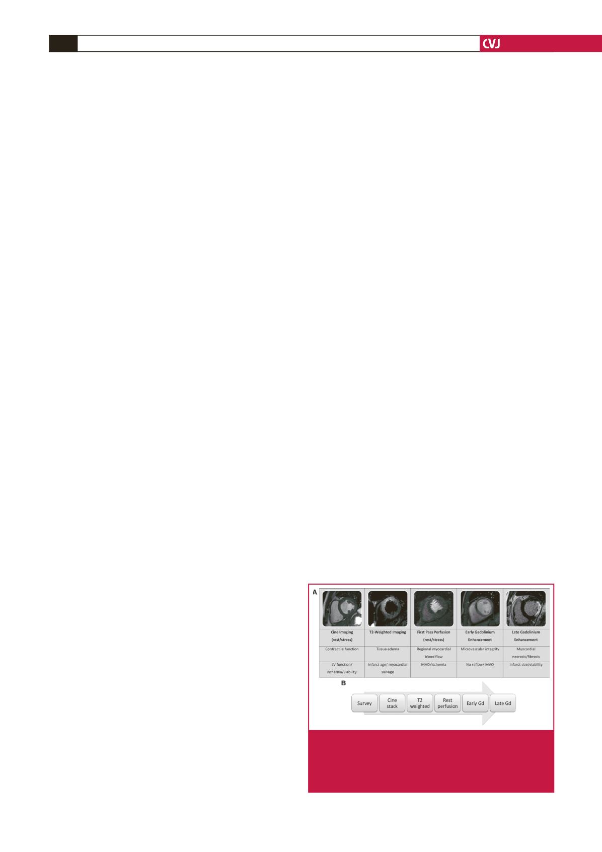
Fig. 1.
CMR methods for the assessment of ACS. Short-axis
views of different patients illustrate the different imag-
ing techniques used (rows 1 and 2), their morpho-
logical correlates (row 3), and main clinical application
(row 4).


