
CARDIOVASCULAR JOURNAL OF AFRICA • Volume 31, No 6, November/December 2020
336
AFRICA
the performing physician and the technician wore sterile single-
use laboratory coats.
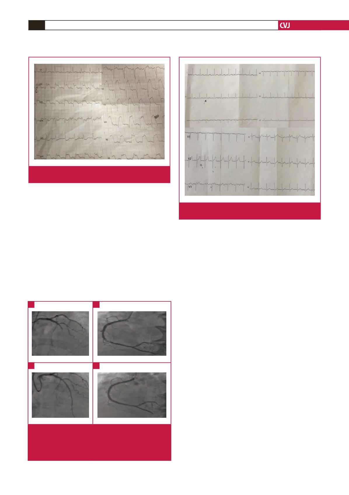
On coronary angiography, the stent in the mid LADwas found
to be patent, however it was occluded totally after the stent, and
the right coronary artery (RCA) had subtotal occlusion. Initially,
a Partner sirolimus-eluting stent of 2.75 × 15 mm was placed in
the mid LAD (Fig. 2). After stent implantation, some blood flow
was observed beyond the occlusion, however, a thrombotic total
occlusion occurred in the distal LAD. Repeated dilatations were
performed on this site, however no blood flow was observed.
Two Boston Scientific Rebel bare-metal stents of 4.0 × 24 mm
and 4.0 × 16 mm were directly implanted (Fig. 2) into the RCA.
In our hospital we have two catheterisation units. The
laboratory used for the intervention was sterilised with
disinfectant solutions and ultraviolet light and closed for 48
hours. The patient was taken into the intensive care unit. After the
intervention, we observed resolution of ST-segment elevations
on his ECG (Fig. 3). On a transthoracic echocardiogram (TTE),
the ejection fraction was observed at 45% with apical akinesia.
In his control TTE performed on 6 April, we observed similar
findings after the intervention. The patient was discharged as
healthy on 21 April.
Discussion
COVID-19 is a pandemic spreading over approximately 208
countries/regions. By 6 April 2020, more than 1 287 742 patients
had become infected worldwide and it had caused the death of
more than 70 000 people.
The prevalence of CVD is higher among those with COVID-
19, and myocardial injury occurs in more than 7% of patients
due to the infection (22% of them critically ill).
3
COVID-19
infection can directly affect the cardiovascular system and the
presence of CVD also facilitates COVID-19 infection.
4
The
Chinese Centre for Disease Control and Prevention reported
in the recently published largest case series in mainland China
that the overall mortality rate was 2.3% (1 023 deaths in 44 672
confirmed cases); however, the mortality rate increased up to
10.5% in patients with a history of CVD.
5
STEMI is still a significant underlying factor for increased
morbidity and mortality rates all around the world, although
there has been a decrease in incidence and an increase in survival
rates recently.
6
Thrombotic occlusion occurs in a coronary
artery at the site of a ruptured or eroded plaque and it leads
to STEMI.
6
Characteristic symptoms, and changes in the ECG
are the basis for diagnosis, and elevated cardiac enzymes
subsequently confirm the diagnosis.
6
If it is performed by
experienced specialists timeously, mechanical reperfusion via
PPCI is superior to fibrinolytic therapy.
6
Fig. 1.
Electrocardiography of the patient showing inferior and
anteroseptal myocardial infarction.
Fig. 3.
Electrocardiography of the patient after primary percu-
taneous coronary intervention.
Fig. 2.
A, C. Angiographic views of the left anterior descending
artery before and after primary percutaneous coronary
intervention. B, D. Angiographic views of the right
coronary artery before and after primary percutaneous
coronary intervention.
A
C
B
D


