CARDIOVASCULAR JOURNAL OF AFRICA • Vol 21, No 2, March/April 2010
110
AFRICA
despite optimised medical and electrical therapy, the patient was
evaluated for cardiac transplantation. A right heart catheterisa-
tion revealed a PA pressure of 68/38 mmHg with a mean of
49 mmHg and pulmonary capillary wedge pressures of 41/64
mmHg with a mean of 49 mmHg, and a cardiac output of 1.63 l/
min with a cardiac index of 0.89 l/min/m
2
.
Continuous intravenous (IV) inotropic therapy using
dobutamine was initiated at 5 mcg/kg/min in combination with
IV diuretics. The cardiac index improved to 1.8 l/min/m
2
, and
with persistent symptoms of heart failure after completing the
evaluation process, the patient was listed for cardiac transplanta-
tion as a status IB.
Even before dobutamine therapy, the patient had recurrent
episodes of ventricular tachycardia, supraventricular tachycardia
and intermittent atrial fribrillation. Amiodarone therapy was
initiated with an initial bolus of 150 mg IV, followed by continu-
ous infusion of 1 mg/min for six hours, followed by 0.5 mg/min
for 18 hours as a continuous infusion, and then changed to oral
amiodarone 200 mg given twice daily 12 hours apart.
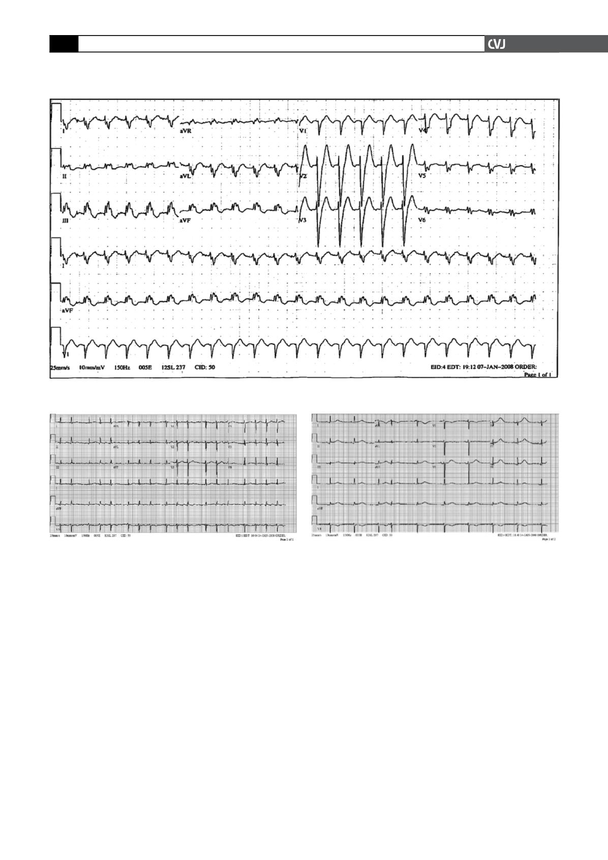
The patient’s rhythm stabilised (Fig. 1) and after 10 days,
orthotopic cardiac transplantation was performed successfully.
The donor heart was from a 36-year-old male without any prior
cardiovascular history or prior anti-arrhythmic therapy. The
donor’s ECG was without any abnormalities.
On postoperative day seven, the recipient developed parox-
ysmal atrial fibrillation (Fig. 2). A single amiodarone bolus of
150 mg was given, followed by an infusion of 0.5 mg/min and
the patient converted to sinus rhythm. Subsequently, the patient
demonstrated prolonged QT interval on ECG (550 msec, QTc
544 msec, Fig. 3) and developed torsade de pointes, which
degenerated into ventricular fibrillation, with successful external
defibrillation.
Amiodarone was discontinued, electrolytes were in the normal
range but IV magnesium was started for a borderline low magne-
sium level of 1.5 mg/dl. Despite shorter but still prolonged QT
intervals the following day (454 ms, QTc 517 msec) with normal
Fig. 1. ECG 1 of the patient’s heart directly prior to surgery (January 4), after treatment with amiodarone, showing
sinus tachycardia with a rate of 134/min, right axis deviation and left bundle branch block. QT duration is 346 msec.
Fig. 2. ECG 2 of the same patient seven days after trans-
plantation (January 11) of the donor heart, showing
paroxysmal atrial fibrillation and heart rate approximately
105/min. QT duration is 344 msec.
Fig. 3. ECG 3 of the recipient after cardiac transplantation
(January 13) with sinus rhythm, heart rate 60/min and
prolonged QT interval of 550 msec. Shortly thereafter, the
patient developed ventricular fibrillation.


