CARDIOVASCULAR JOURNAL OF AFRICA • Vol 21, No 2, March/April 2010
114
AFRICA
References
Dursun M,Yilmaz S, Sayin OA, Ugurlucan M, Ucar A,Yekeler E, Tunaci
1.
A. Combination of unicuspid aortic valve, aortic coarctation, and aber-
rant right subclavian artery in a child: MR imaging and CTA findings.
Cardiovasc Intervent Radiol
2007;
30
: 547–549.
Novaro GM, Mishra M, Griffin BP. Incidence and echocardiographic
2.
features of congenital unicuspid aortic valve in an adult population.
J
Heart Valve Dis
2003;
12
: 674–678.
Falcone MW, Roberts WC, Morrow AG, Perloff JK. Congenital aortic
3.
stenosis resulting from a unicommisssural valve. Clinical and anatomic
features in twenty-one adult patients.
Circulation
1971;
44
: 272–280.
Roberts WC, Ko JM. Clinical and morphologic features of the congeni-
4.
tally unicuspid acommissural stenotic and regurgitant aortic valve.
Cardiology
2007;
108
: 79–81.
Collins MJ, Butany J, Borger MA, Strauss BH, David TE.
5.
Implications
of a congenitally abnormal valve: a study of 1025 consecutively excised
aortic valves.
J Clin Pathol
2008;
61
: 530–536.
Singh D, Chee TS. Incidental diagnosis of unicuspid aortic valve in an
6.
asymptomatic adult.
J Am Soc Echocardiogr
2008;
21
: 876.e5.
Murphy BA, Groban L, Kon ND. Diagnosis of a unicuspid aortic valve
7.
using transesophageal echocardiography.
J Cardiothorac Vasc Anesth
2003;
17
: 82–83.
Ishigami H, Iwase M, Hyoudo K, Aoyama I, Ito M, Tajima K,
8.
et al
. A
case of unicuspid aortic valve associated with a single coronary artery
and ventricular septal defect.
J Med Ultrason
2005;
32
: 65–70.
Schäfers HJ, Aicher D, Riodionycheva S, Lindinger A, Rädle-Hurst T,
9.
Langer F, Abdul-Khaliq H. Bicuspidization of the unicuspid aortic valve:
a new reconstructive approach.
Ann Thorac Surg
2008;
85
: 2012–2018.
Bansal A, Arora S, Traub D, Haybron D. Unicuspid aortic valve
10.
and aortic arch aneurysm in a patient with Turner syndrome.
Asian
Cardiovasc Thorac Ann
2008;
16
: 266–267.
Debl K, Djavidani B, Buchner S, Poschenrieder F, Heinicke N, Schmid
11.
C,
et al
. Unicuspid aortic valve disease: a magnetic resonance imaging
study.
Rofo
2008;
180
: 983–987.
Gibbs WN, Hamman BL, Roberts WC, Schussler JM. Diagnosis of
12.
congenital unicuspid aortic valve by 64-slice cardiac computed tomog-
raphy.
Proc (Bayl Univ Med Cent)
2008;
21
: 139.
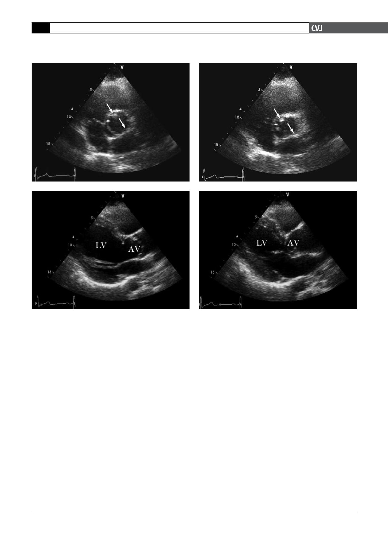
Fig. 1. Transthoracic echocardiaography showing a unicuspid aortic valve with a raphe at the 11 o’clock position
(upper arrow) and a clear commissure at the 4–5 o’clock position (lower arrow) on a short-axis view during systole
(A), and diastole (B). The aortic valve in an integral movement and in a dome-shaped configuration during systole (C)
and diastole (D), and left ventricular hypertrophy and dilated aortic root extending 3.8 cm in diameter could be seen
from the parasternal long axis view (C, D). AV: aortic valve; LV: left ventricle.
A
B
C
D


