CARDIOVASCULAR JOURNAL OF AFRICA • Vol 22, No 1, January/February 2011
36
AFRICA
Coronary artery ectasia in a patient with myocardial
infarction
ML JESURAJ, D MUKERJEE, AV JESURAJ, R SINGH, B AGARWAL
Summary
We report on a case of triple-vessel coronary artery ecta-
sia (CAE) in a young patient. This patient presented with
anterior wall myocardial infarction (MI) with post-infarct
angina. His coronary angiogram revealed coronary artery
ectasia involving the left anterior descending, circumflex and
right coronary arteries.
Keywords:
coronary artery ectasia, myocardial infarction
Submitted 23/3/09, accepted 14/3/10
Cardiovasc J Afr
2011;
22
: 36–37
DOI: CVJ-21.019
Coronary artery ectasia (CAE) is an uncommon disorder
diagnosed in one to 4% of patients undergoing coronary arte-
riography.
1-3
CAE is usually considered a variant of athero-
sclerotic coronary artery disease.
1-3
Coronary artery disease in
young adults usually occurs in patients with multiple predis-
posing factors, such as hyperlipidaemia, cigarette smoking,
diabetes mellitus, hypertension, and a strong family history.
In the absence of predisposing factors, other causes should be
considered, particularly mucocutaneous lymph node syndrome
[Kawasaki disease (KD)].
4
Case report
A 36-year-old man presented with a history of acute anterior
wall myocardial infarction (MI) with post-infarct angina. He was
stabilised with medical therapy. There was no history of hyper-
tension, diabetes mellitus or hyperlipidaemia. The patient was a
non-smoker. He was unable to recall any definite symptoms of
acute KD in childhood. The physical examination and laboratory
data disclosed no abnormalities. The lipid profile, homocysteine
and lipoprotein (a) [Lp(a)] levels were within normal limits.
An ECG showed features of recent extensive anterior wall
MI. A two-dimensional Doppler echocardiogram showed a left
ventricular ejection fraction of 46% with anterolateral wall
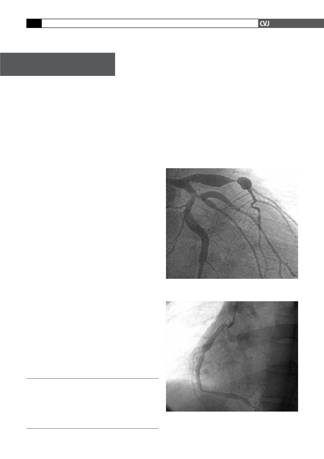
hypokinesis. Coronary arteriography (Figs 1, 2) demonstrated
severe disease involving the left anterior descending, the circum-
flex and the right coronary arteries. The proximal segments of
the arteries were very ectatic. Multiple aneurysms alternating
with severe stenoses were seen along the entire length of the
vessels, an appearance typically seen in KD.
4
He was stabilised
Case Report
Institute of Cardiovascular Sciences, IPGME&R, Kolkata,
West Bengal, India
ML JESURAJ, MD,
D MUKERJEE, MD
R SINGH, MD
B AGARWAL, MD
Department of Pathology, VIMS, Kolkata, West Bengal, India
AV JESURAJ, MD
Fig. 1. Selective left coronary angiogram demonstrating
severe ectasia of the proximal segments of the left coro-
nary artery with stenotic lesions.
Fig. 2. Selective right coronary angiogram showing multi-
ple ectatic segments and stenosis of proximal and mid
segments.


