CARDIOVASCULAR JOURNAL OF AFRICA • Vol 22, No 5, September/October 2011
AFRICA
247
root, MVP, MR or dilatation of the pulmonary artery. Despite
the diffuse abnormality of the aortic wall, enlargement usually
occurs at the sinuses of Valsalva. This condition is seen in 70 to
80% of MFS patients and is more common in males. Dilatation
of the aortic root usually begins in childhood. Independent of
age, the rate of progression of the enlargement increases after the
aortic diameter exceeds 5 cm. At this stage, the risk of dissection
and rupture of the ascending aorta increases and therefore surgi-
cal intervention is suggested. Progressive dilatation of the aortic
root also impairs leaflet coaptation and this causes AR.
1–3
Mitral valve disease is present in 60 to 80% of the MFS popu-
lation. This usually occurs in childhood before aortic involve-
ment. Common mitral valve abnormalities are annular dilatation,
fibromyxomatous changes in leaflet and chordae, elongated
chordae, rupture of the leaflets and calcium deposition. Proximal
pulmonary artery enlargement in the absence of pulmonary
valve involvement or peripheral pulmonary artery stenosis, TVP,
and coronary, axillary and subclavian artery aneurysms may also
be seen in MFS.
1-3
Among the patients evaluated for marfanoid phenotype, 11
were diagnosed as MFS. Although none had cardiac symptoms,
dilatation of the aortic root was found in six patients and the
diagnosis of MFS was based on cardiac involvement in this
group of patients.
3
Five others with valvular abnormalities with-
out dilatation of the aortic root were diagnosed with the help of
findings in other systems. These valvular abnormalities were
accepted as minor findings, according to the Ghent criteria.
3
Despite the fact that prognosis of patients with MFS depends
on the presence of dilatation of the aortic root, there is insuf-
ficient information on aortic root measurements in children.
There is consensus on measuring aortic root dimensions at
the level of the sinuses of Valsalva but no agreement on which
echocardiographic technique to use, cross-sectional or M-mode
echocardiography. In our study, measurements were made from
both cross-sectional and M-mode views and mean values of both
measurements were used.
9-12
Diagnosis from dilatation of the aortic root was based on the
monogram defined by Roman and colleagues.
8
Because aortic
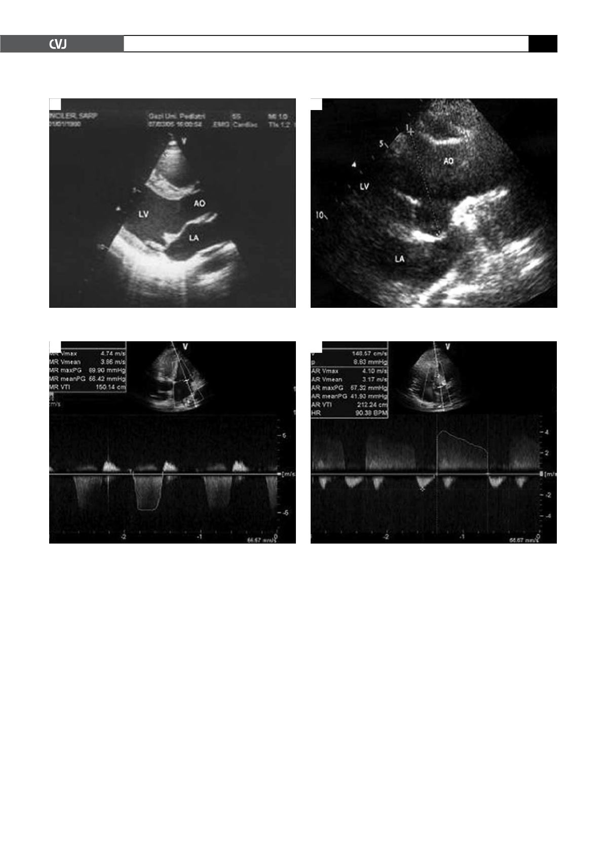
Fig. 2. Mitral valve prolapse (A), and dilatation of the aortic root (B) in cross-sectional echocardiographic examination.
AO: aorta, LA: left atrium, LV: left ventricle.
A
B
Fig. 3. Mitral valve regurgitation in apical four-chamber view (A), and aortic regurgitation in apical five-chamber view
(B) with pulse-wave Doppler.
A
B


