
CARDIOVASCULAR JOURNAL OF AFRICA • Volume 26, No 6, November/December 2015
e2
AFRICA
crepitus on palpation around the sternal notch. Auscultation
of the heart revealed a loud crunch-like sound during systole
consistent with Hamman sign. Neurological and abdominal
examinations showed no abnormalities.
Laboratory tests, including cardiac enzymes and an
electrocardiogram, were normal. A postero-anterior chest
radiograph was normal, but a chest computed tomography
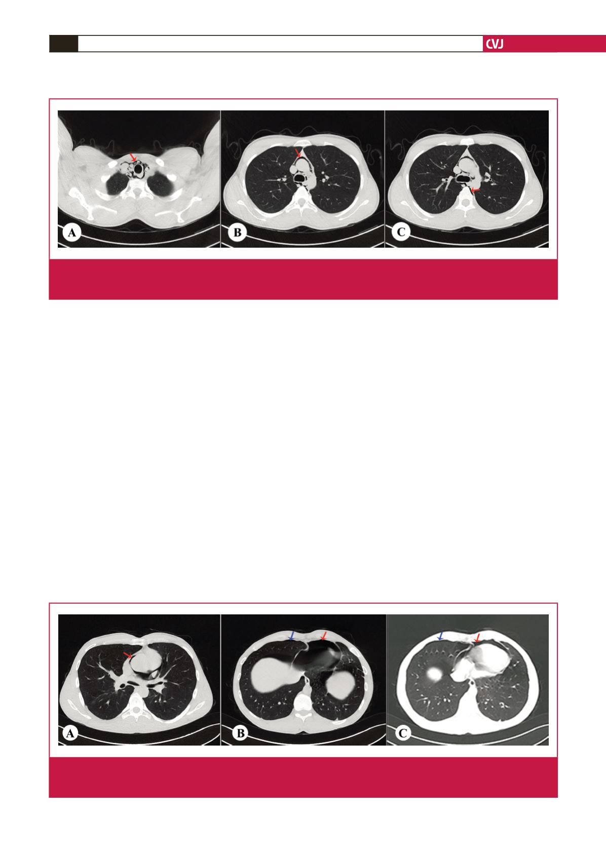
(CT) scan showed subcutaneous emphysema and pneumo-
mediastinum (Fig. 1). There was no evidence of pneumothorax,
pneumopericardium, pulmonary parenchymal injury, rib
fractures, or tracheal or bronchial injuries.
The patient was transferred to the thoracic surgery department
and admitted to hospital for observation and non-surgical
treatment. His progress was uneventful and he was discharged
after four days. Written informed consent was obtained from the
patient for the publication of this case report.
Case 2
A 23-year-old man was admitted to the emergency department
because of a two-day history of dyspnoea and chest pain.
He had no history of trauma. The blood pressure was 120/85
mmHg, pulse was 91 beats per minute, respiratory rate was 18
breaths per minute, temperature was 37°C, and transcutaneous
oxygen saturation was 93% on room air. There was tenderness to
palpation in the right hemithorax and around the sternum. The
breath sounds were normal and equal in both lungs.
Laboratory tests, including cardiac enzymes and an
electrocardiogram, were normal. The postero-anterior chest
radiograph showed a right pneumothorax and transparency
that was consistent with left mediastinal air. Thoracic CT scan
showed right pneumothorax and pneumomediastinum (Fig. 2).
The patient was transferred to the thoracic surgery department
and admitted to hospital for observation and non-surgical
treatment. His progress was uneventful and he was discharged
after five days. Written informed consent was obtained from the
patient for the publication of this case report.
Discussion
The chief complaint on presentation to the emergency
department in both patients included chest pain and dyspnoea.
The first patient had traumatic pneumomediastinum as a result
of blunt chest trauma, which is a rare clinical condition. The
second patient had spontaneous pneumomediastinum. In this
study, we investigated the diagnosis and treatment of the two
Fig. 1.
Case 1: axial thoracic computed tomography showing free air density consistent with pneumomediastinum (A) around the
trachea in the upper mediastinum, (B) around the aorta in the lower mediastinum, and (C) at the posterior oesophagus (red
arrows).
Fig. 2.
Case 2: axial thoracic computed tomography showing free air density consistent with pneumomediastinum (A) around the
aorta and pulmonary artery, (B) inferior to the heart, and (C) anterior to the heart (red arrows). In addition, pneumothorax
was detected (B and C) in the anterior right hemithorax (blue arrows).


