


 36 / 64

CARDIOVASCULAR JOURNAL OF AFRICA • Volume 27, No 1, January/February 2016
34
AFRICA
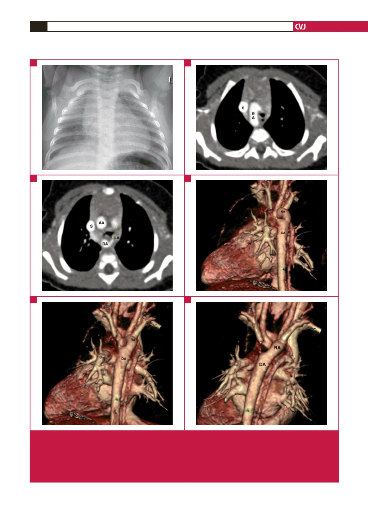
Fig. 4.
Double aortic arch with an atretic left aortic arch. (A) The plain chest radiograph suggests a right aortic arch, with a widened
right paratracheal shadow. (B) and (C) The axial CTA images confirm the dominant RA, with the non-dominant LA consist-
ing of a short patent segment near the DA and a longer atretic segment (between the yellow asterisks). The LA, together
with the left ligamentum arteriosum, completes the vascular ring encircling the aerodigestive tract (red asterisk). (D), (E),
(F) Postero-anterior oblique CTA reconstruction views illustrate the position of the atretic and thus ‘invisible’ left aortic arch
(between the white asterisks), thereby completing the ring. RA: right arch; LA: left arch; S: superior vena cava; DA: descend-
ing aorta; AA: ascending aorta.
A
E
C
B
F
D