


 39 / 102

CARDIOVASCULAR JOURNAL OF AFRICA • Volume 27, No 3, May/June 2016
AFRICA
161
whereas severe endothelial injury and tunica media defects were
seen in the control group.
Within the frame of classification of endothelial injury, in
the control group, no significant injury was observed in three
samples, whereas type 1, type 2 and type 3 vascular endothelial
injury was seen in six, six and five grafts, respectively. In the
cyanoacrylate group, no endothelial injury was observed in
seven grafts, type 1 and type 2 endothelial injury was seen in
10 and two grafts, respectively, and there was no type 3 injury
in the grafts. Endothelial injury was significantly less in the
cyanoacrylate group, as shown by the assessment of intergroup
results (Table 1).
The cyanoacrylate group did not exhibit any significant
change in the medial layer of the saphenous vein grafts, whereas
Masson’s Trichrom staining demonstrated significant separation
and oedema between the tunica media and intima in the
untreated saphenous vein graft group.
Discussion
In this study, a model of the arterial system was established
and the saphenous vein graft was exposed to internal pressure.
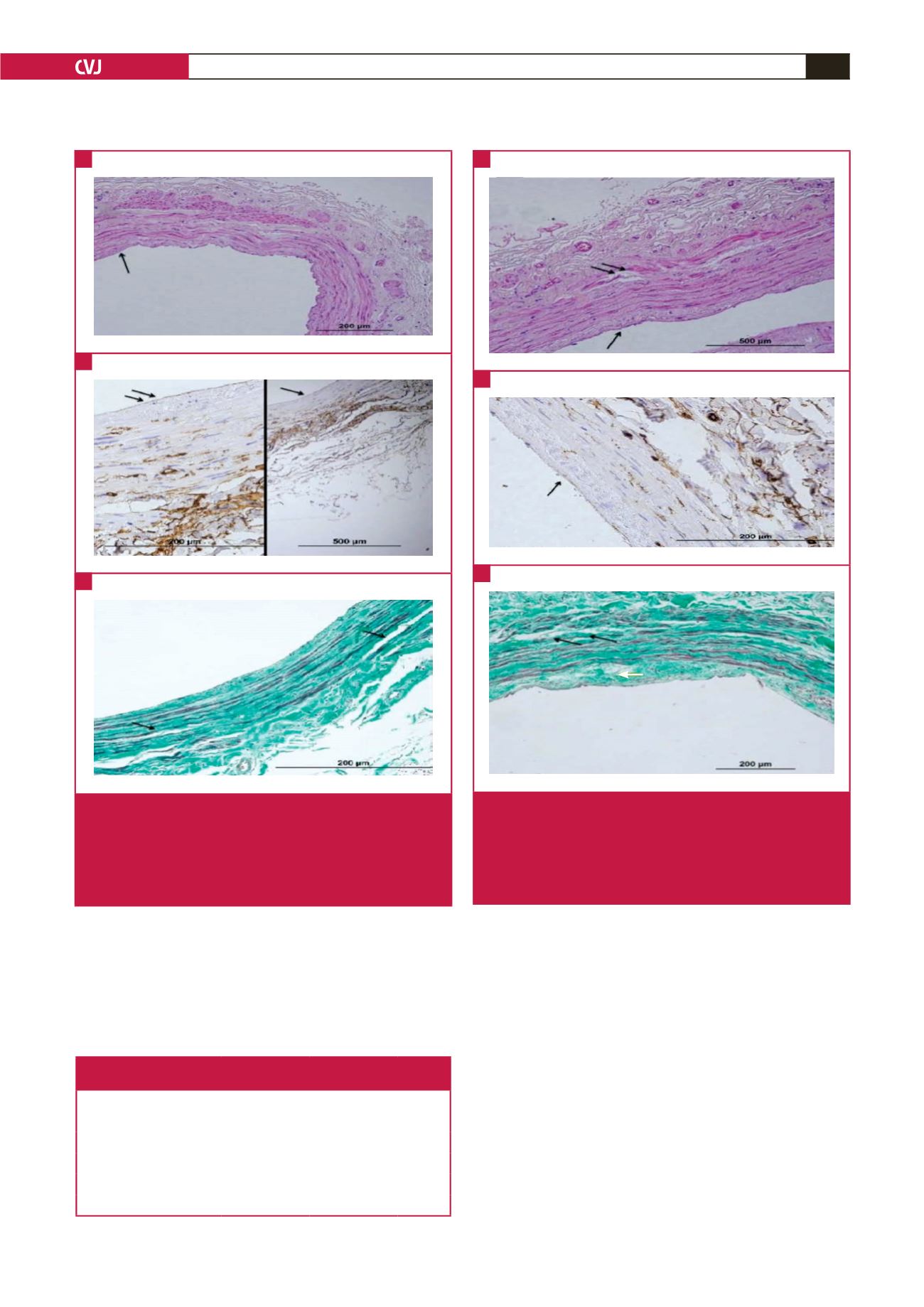
Fig. 3.
A shows moderate endothelial cell loss (black arrows).
B shows moderate loss of CD34-labelled endothelial
cells (black arrows). C shows significant separation
between the tunica media and intima (black arrows),
and oedema (white arrow). Additionally, there is mild
loss in the organelle distribution in the collagen fibres.
A
B
C
Fig. 4.
A shows severe endothelial cell loss (black arrows). B
shows nearly total loss of CD34-labelled endothelial
cells (black arrows). C shows significant separation
between the tunica media and intima (black arrows),
and oedema (white arrow). Additionally, there is loss in
the organelle distribution in the collagen fibres.
A
B
C
Table 1. Distribution of saphenous vein injury
in the groups as per classification
Class of vascular damage
Group 1
(perivascular
cyanoacrylate)
Group 2
(control)
p
-value
No injury
7
3
0.003
Type 1 injury
10
6
Type 2 injury
2
6
Type 3 injury
0
5