


 38 / 102

CARDIOVASCULAR JOURNAL OF AFRICA • Volume 27, No 3, May/June 2016
160
AFRICA
line of the pump (Fig. 1). Cyanoacrylate was sprayed on the
outer surface of one of the saphenous vein grafts and allowed
to dry for five minutes. The other saphenous vein graft was
not subjected to any process and both grafts were exposed to
pump flow at 120 mmHg for approximately 45 minutes. At the
end of the procedure, both saphenous veins were placed into
containers with 10% formaldehyde solution and dispatched to
the laboratory for histopathological examination.
Histopathological examination
The removed grafts were fixed in 10% buffered formaldehyde
solution for 24hours.After routine tissue processing, 5-
µ
msections
were cut from the paraffin-embedded blocks. These sections were
stained with haematoxylin and eosin (H&E), histochemically
with Masson’s Trichrom, and immunohistochemically with
CD34, which is an endothelial marker. All sections were coded
and the endothelium was examined under a light microscope by
a pathologist who was unaware of the treatment protocol applied
(Olympus CX 51, Tokyo, Japan).
The histomorphological classification of endothelial injury
was as follows:
•
No injury: endothelial cells are in contact with each other,
the cell has no change in contents or reduction in diameter.
Platelets and other blood cells may or may not have adhesions
to the endothelium (Fig. 2).
•
Type 1 injury: while the integrity of endothelial cells is main-
tained on the entire endothelial surface and endothelial cells
are in contact with each other, there is a change in contents
and reduction in diameter (flattening) of the cell. There is
adhesion of the platelets and other blood cells to the endothe-
lium (Fig 2).
•
Type 2 injury: there is detachment of the cells from the junc-
tions and lack of endothelial cells in places (Fig. 3).
•
Type 3 injury: there is endothelial cell peeling and the subse-
quent formation of sub-endothelial tissue (Fig. 4).
Statistical analysis
The variables obtained were classified into categories and
indicated as numbers and percentages. The chi-squared test
was used to evaluate the analysis of categorical data. SPSS 18
(SPSS Inc, Chicago, IL, USA) software was used for statistical
evaluations.
Results
Endothelial injury was determined from the diameter of the
saphenous vein, which remained unchanged in the group
supported with cyanoacrylate, whereas severe distention of the
saphenous vein occurred in the control group.
Endothelial injury was examined by H&E staining and
immunohistochemically with CD34 staining, which is an
endothelial marker. On first impression, no severe damage was
seen in the saphenous vein grafts from the cyanoacrylate group,
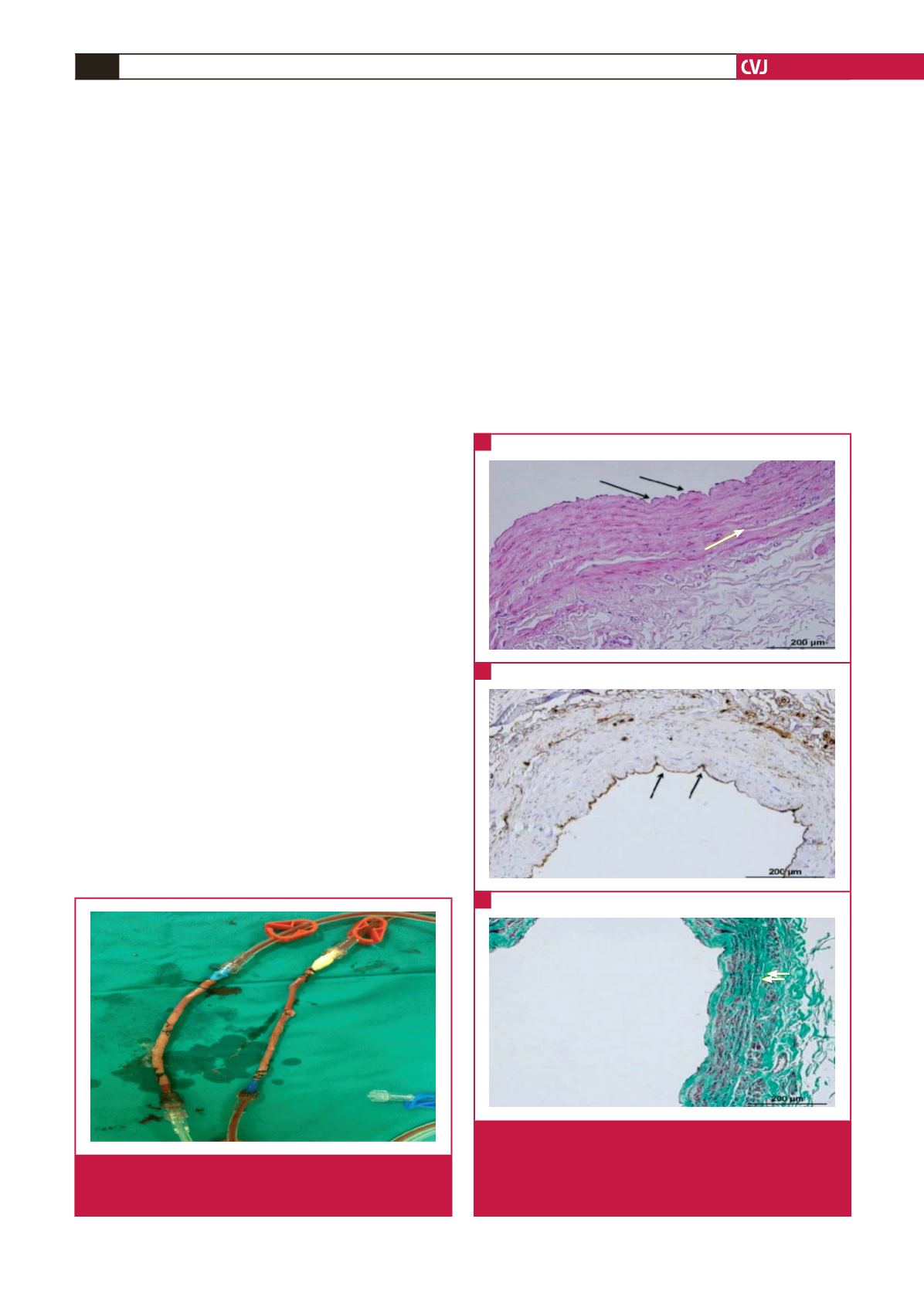
Fig. 1.
Perivascular cyanoacrylate applied to one of the saphe-
nous vein grafts, prepared from the same patient, for
the purpose of external support.
Fig. 2.
A shows mild endothelial cell loss (black arrows) with
no oedema and no minimal intimal separation (white
arrow). B shows CD34-labelled endothelial cell loss
(black arrows). C shows minimal separation of the
tunica media and intima but no loss of organelles.
A
B
C