
CARDIOVASCULAR JOURNAL OF AFRICA • Volume 28, No 2, March/April 2017
82
AFRICA
years) who had had coronary catheterisation performed by
interventional cardiologists for symptoms suggestive of CAD. In
order to assess the effect of CACs on LV function in the presence
of total occlusion of the coronary artery, only those coronary
angiograms that had LV function assessed by ventriculography
were selected for analysis.
Ninety-seven such patients with total occlusion of a coronary
artery and LV functional assessment were included in the
analysed angiograms. The mean EF and the different grades of
CACs in these patients were determined.
The angiograms were obtained from the cardiac
catheterisation laboratories of hospitals within the private sector
in the eThekwini municipality region of KwaZulu-Natal, South
Africa. Ethical approval (ethics number BE 196/13) for the study
was obtained from the University of KwaZulu-Natal Biomedical
Research Ethics Committee.
Coronary arteriography was performed via the percutaneous
transfemoral approach by injecting a radio-opaque contrast agent
into the coronary blood vessels, and the images were taken using
X-ray fluoroscopy. These images were recorded on digital media
in DICOM (Digital Imaging and Communication in Medicine)
format and stored in the cardiac catheterisation laboratories.
The relationship between the location of the atherosclerotic
lesions and the CAC grades were examined in the angiograms
that met the inclusion criteria. In addition, the relationship
between the location of the atherosclerotic lesions and the mean
EF was also evaluated. The location of atherosclerotic lesion
was determined by dividing the coronary arteries into proximal,
middle and distal regions.
The Rentrop grading system
22
is the most widely used grading
system for coronary collaterals and is employed by many
researchers. However, most patients are graded Rentrop 2 or 3 in
chronic total coronary occlusion.
23
The grading of the coronary collaterals in the present study
was based on the grading system used by Werner
et al
.,
24
with
the addition of a grade for absent CACs. This system centered
on defining the collateral connection between the donor and
the recipient arteries. Therefore, in this study, the coronary
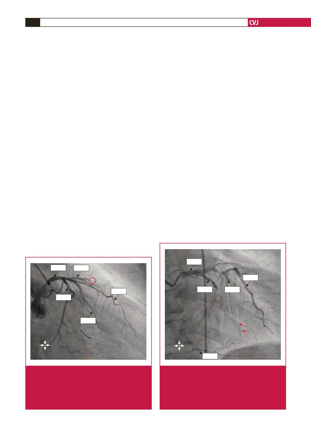
collaterals were graded as: grade 0 for absent collateralisation,
where there were no demonstrable CACs to the distal region of
the obstructed vessel (Fig. 1); grade 1 for poor collateralisation,
where there were CACs showing no continuous connection
between the donor and recipient arteries (Fig. 2); grade 2 for
good collateralisation, where there were continuous threadlike
connections between the donor and recipient arteries; and grade
3 for excellent collateralisation, where there were continuous
prominent connections with side branches between the donor
and recipient arteries (Fig. 3).
Data were analysed with the Statistical Package for the Social
Sciences (SPSS) version 21 for Windows (IBM SPSS, NY, USA).
A
p
-value
<
0.05 was considered statistically significant.
Results
The mean age of the patients with coronary artery occlusion
who had LV function assessed by ventriculography was 59
±
8 years. The patients consisted of 25.8% females and 74.2%
males (Table 1). The grades of the CACs were as follows: absent
(15.4%), poor (15.4%), good (36.9%) and excellent (32.3%). The
morphological properties of the coronary arterial tree in the
analysed angiograms are shown in Table 1.
The grades of the collateral pathways with regard to the
location of atherosclerotic obstruction were evaluated. They
were recorded as 15.9, 9.1, 34.1 and 40.9% in the proximal region
LCA
LAD
D
Cx
OM
R L
S
I
Fig. 1.
Coronary angiogram in the right anterior oblique
view (caudal angulation) showing obstruction of the
diagonal branch of the left anterior descending (LAD)
artery (red ring) without collateral vessels to the distal
segment of the obstructed vessel.
LCA, left coro-
nary artery; D, diagonal; Cx, circumflex; OM, obtuse
marginal artery.
R L
S
I
LAD
LCA
Cx
PDA
SB
Fig. 2.
Coronary angiogram in the right anterior oblique view
showing the filling of the posterior descending artery
of an obstructed right coronary artery (RCA) by grade
1 collateral vessel (red arrows) originating from the
septal branch of the left anterior descending (LAD)
artery. LCA, left coronary artery; Cx, circumflex; SB,
septal branch; PDA, posterior descending artery.


