


 64 / 68

CARDIOVASCULAR JOURNAL OF AFRICA • Volume 31, No 2, March/April 2020
e6
AFRICA
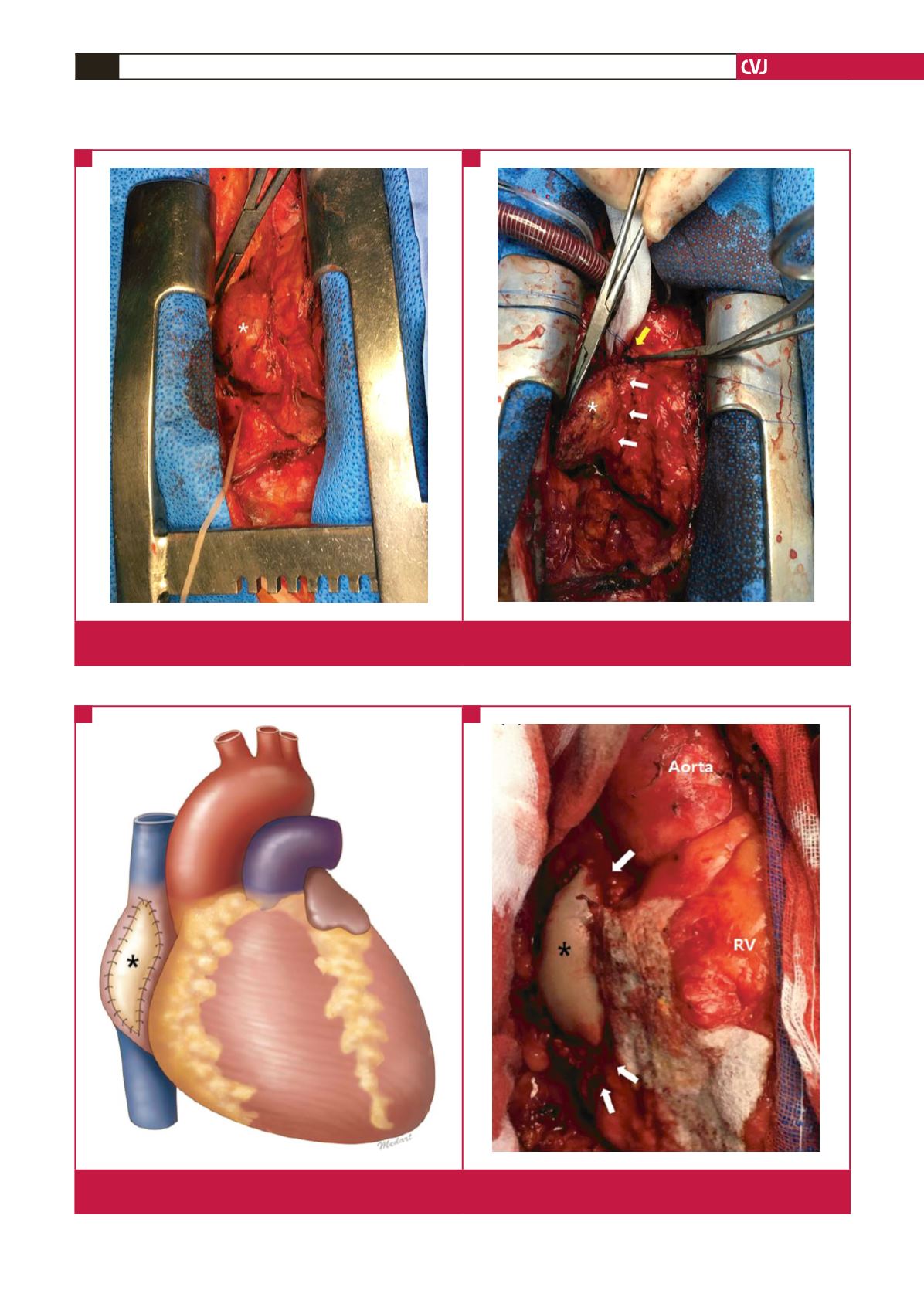
Fig. 1.
(A) A 5-cm ovoid mass (asterisk) was strongly adhered to the right atrium after a median sternotomy. (B) Right atrial injury
(yellow arrow) occurred during dissection of the mass (asterisk). Right atrium is indicated by white arrows.
A
B
Fig. 2.
(A) Schematic illustration of the right atrial reconstruction. (B) Intra-operative photograph. The right atrium (white arrows) was
reconstructed using bovine pericardium (asterisk) after removing the mass. RV, right ventricle.
A
B