CARDIOVASCULAR JOURNAL OF AFRICA • Vol 24, No 7, August 2013
AFRICA
257
(group 1) was higher than in the vitamins C
+
E group (group
2) and the control group. Also, a history of mild hypertension in
the vitamin C group was higher than in the vitamins C
+
E and
control groups but it was not statistically significant.
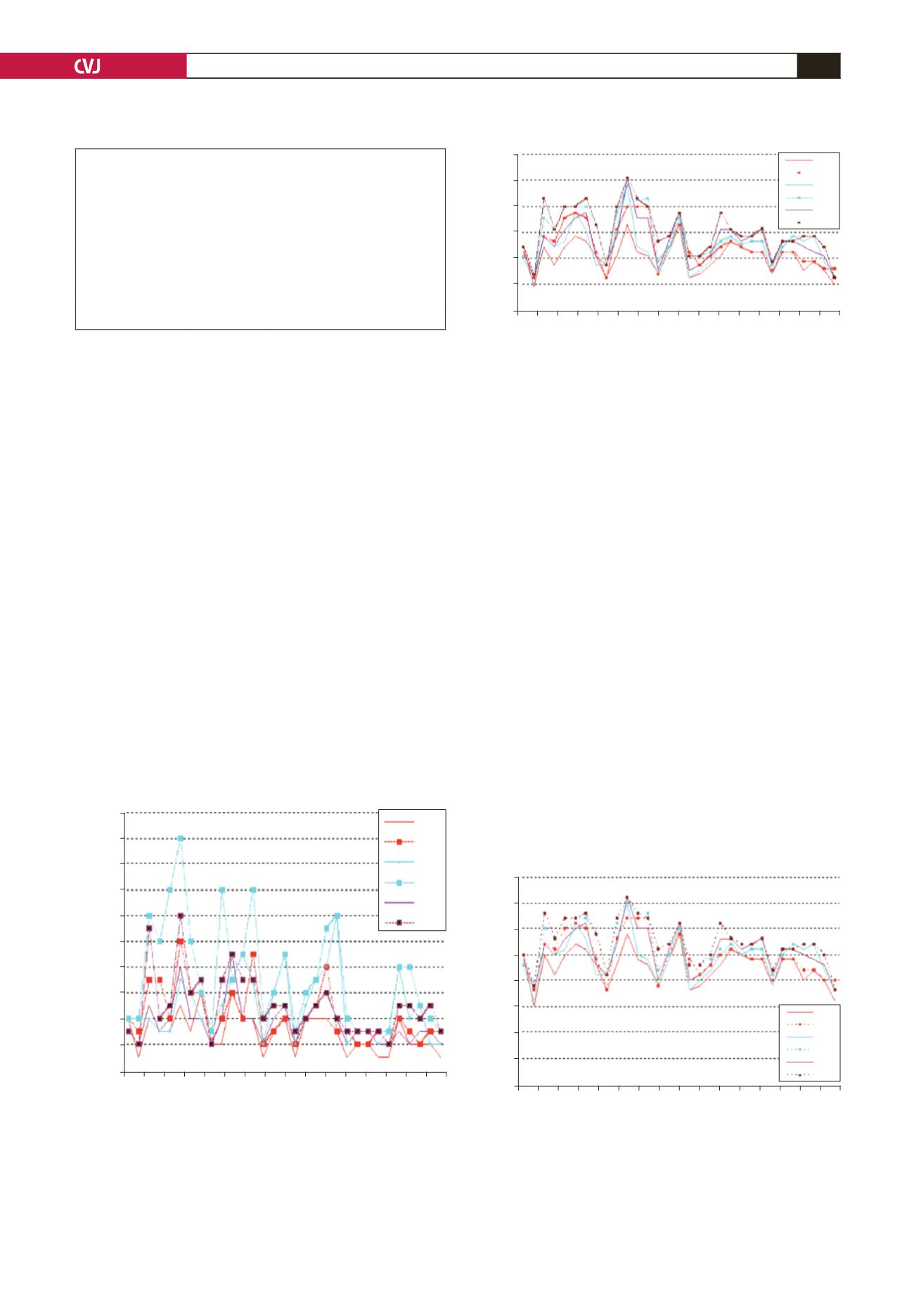
Pre- and post-administration measurements of vitamin C in
group 1 showed a statistically significant increase in the radial
artery flow volume, lumen diameter and lumen area after two
hours of vitamin C administration (Figs 1–3) (
p
<
0.001). Pre-
and post-administration measurements of vitamins C
+
E in
group 2 showed a statistically significant increase in the radial
artery flow volume, lumen diameter and lumen area after two
hours of vitamin C with vitamin E administration (Figs 4–6) (
p
<
0.001).
Patients in both the vitamin C group (measurement 4, 5, 6
for group 1) and in the vitamins C
+
E group (measurement
4, 5, 6 for group 2) showed statistically significant increases
in the radial artery flow volume, lumen diameter and lumen
area when compared with the time of measurements 1, 2 and
3. Its combination with vitamin E was superior to vitamin C
administration alone for endothelium-dependent vasodilatation
but this difference was not statistically significant. This is shown
in Table 2.
Against the two groups, there was no statistical difference in
the control group. The repeat measurements were not statistically
different from the first measurements and they were also not
different from the baseline measurements of the two groups.
Discussion
Antioxidants vitamins C and E improved defective endothelial
function. This has been attributed to an enhancement in the
synthesis or prevention of the breakdown of nitric oxide (NO).
7
NO plays a major role in maintaining normal tonus in the artery.
This study demonstrated that administration of vitamin C or its
combination with vitamin E increased endothelium-dependent
dilatation in patients awaiting coronary artery bypass surgery.
The radial artery is increasingly being used as a conduit
for coronary bypass grafting because of reports of long-term
patency, accessibility and encouraging mid-term results.
8-10
The
choice of a potent vasodilator with minimal side effects appears
to be an important parameter in ensuring the success of radial
artery conduits. Also, imaging by Doppler ultrasound of radial
artery dilatation after drug treatment is easy. Therefore we
particularly used the radial artery for measurements.
A history of smoking is an important parameter that can affect
the results. Its vasoconstrictor effect on the endothelium has
been shown in many studies. In our study, the vitamin C group
TABLE 2. SUMMARY OF THE KEY CHARACTERISTICS
OF THE SUBJECTS
Vitamin C
group
Vitamin C
+
E group
Control group
Age, median
56.32
±
12.50 55.74
±
13.36 56.20
±
12.40
Ejection fraction
49.64
±
12.57 51.90
±
9.83 50.30
±
10.47
Male/female
24/7
25/6
26/5
History of smoking
16
11
10
Diabetes mellitus
7
7
4
Mild hypertension
15
10
10
Fig. 1. Flow volume measurements on the radial artery
before and after oral vitamin C administration. FV1, FV4,
baseline flow volume before and after vitamin C, respec-
tively; FV2, FV5, flow volume at the moment of cuff defla-
tion before and after vitamin C, respectively; FV3, FV6,
flow volume 60 seconds after cuff deflation before and
after vitamin C, respectively.
0.2
0.18
0.16
0.14
0.12
0.1
0.08
0.06
0.04
0.02
0
1
3 5 7 9 11 13 15 17 19 21 23 25 27 29 31
Patients
Flow volume (l/min)
FV1
FV4
FV2
FV5
FV3
FV6
Fig. 2. Radial artery area measurements before and
after oral vitamin C administration. A1, A4, baseline area
before and after vitamin C, respectively; A2, A5, area at
the moment of cuff deflation before and after vitamin C,
respectively; A3, A6, area 60 seconds after cuff deflation
before and after vitamin C, respectively.
12
10
8
6
4
2
0
1
3 5 7 9 11 13 15 17 19 21 23 25 27 29 31
Patients
Radial artery area (mm
2
)
A1
A4
A2
A5
A3
A6
Fig. 3. Radial artery diameter before and after oral vita-
min C administration. D1, D4, baseline diameter before
and after vitamin C, respectively; D2, D5, diameter at
the moment of cuff deflation before and after vitamin
C, respectively; D3, D6, diameter 60 seconds after cuff
deflation before and after vitamin C, respectively.
4
3.5
3
2.5
2
1.5
1
0.5
0
1
3 5 7 9 11 13 15 17 19 21 23 25 27 29 31
Patients
Radial artery diameter (mm)
D1
D4
D2
D5
D3
D6


