CARDIOVASCULAR JOURNAL OF AFRICA • Volume 25, No 4, July/August 2014
AFRICA
e1
Case Report
Ross procedure in a child with Aspergillus endocarditis
and bicuspid aortic valve
Fotios A Mitropoulos, Meletios A Kanakis, Constantinos Contrafouris, Cleo Laskari, Spyridon Rammos,
Christos Apostolidis, Prodromos Azariadis, Andrew C Chatzis
Abstract
The case is presented of a previously healthy infant with
a known asymptomatic bicuspid aortic valve who devel-
oped fungal endocarditis. The patient underwent aortic root
replacement with a pulmonary autograft (Ross procedure).
Cultured operative material revealed
Aspergillus
infection.
The patient had an excellent recovery and remained well one
year later.
Submitted 7/4/13, accepted 28/4/14
Cardiovasc J Afr
2014;
25
: e1–e3
DOI: 10.5830/CVJA-2014-031
Case report
A previously healthy 20-month-old girl with a known
asymptomatic bicuspid aortic valve presented to a children’s
hospital with a three-day history of low-grade fever, anorexia,
weight loss and irritability. In spite of the absence of positive
blood cultures, the provisional diagnosis of endocarditis was
made based on clinical history, the presence of a new cardiac
murmur, elevated white blood cell (WBC) count and C-reactive
protein (CRP) level, as well as echocardiographic evidence of
aortic and mitral regurgitation in conjunction with suspicious
vegetations on the aforementioned valves. The patient was put
on broad-spectrum antibiotics and transferred to our hospital
for further management five days post her initial admission.
On arrival, the patient was comfortable and apyrexial. Blood
pressure measured 110/40 mmHg and heart rate 135 beats/min.
There were no endocarditis stigmata. On auscultation, a reduced
second sound was noted, along with a 3/6 ejection systolic and
1/6 diastolic murmur, best heard at the aortic position.
Laboratory investigations showed WBC 16 000 cells/ml,
haemoglobin 8.7 g/dl and CRP 23 mg/l. Echocardiography
revealed mild aortic stenosis (PG 15 mmHg), severe aortic
(3+/4+) and less severe (2+/4+) mitral regurgitation, and left
atrial (LA) and ventricular (LV) enlargement with preserved LV
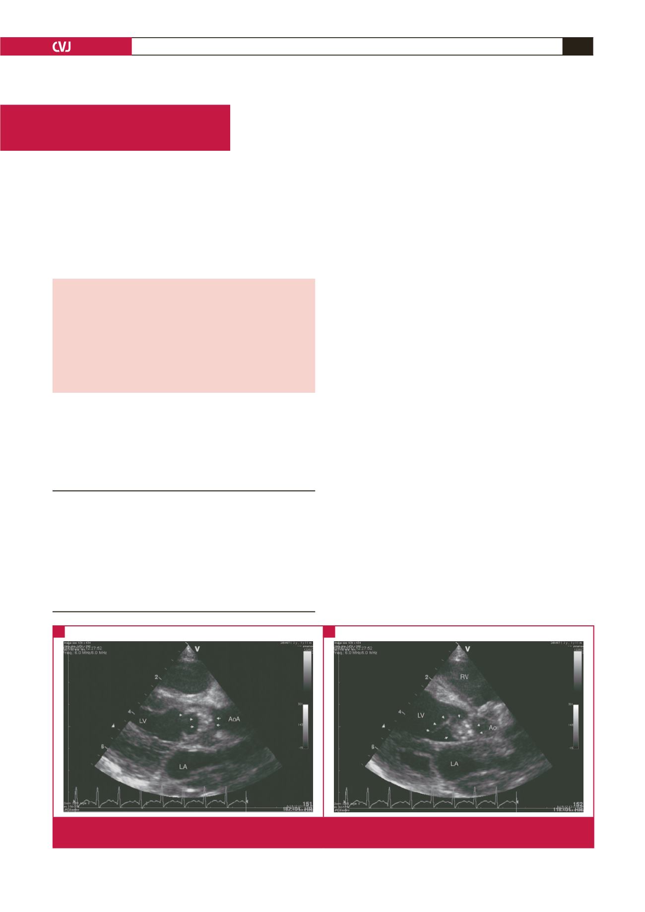
function. Vegetations were noted on both valves (Fig. 1).
Brain, chest and abdominal CT scans were negative for septic
emboli. Repeat blood cultures turned out negative. Since the
haemodynamic burden was well tolerated, the patient continued
Department of Paediatric and Congenital Cardiac Surgery,
Onassis Cardiac Surgery Centre, Athens, Greece
Fotios A Mitropoulos, MD
Meletios Kanakis MD, MD,
Constantinos Contrafouris,
Cleo Laskari, MD
Spyridon Rammos, MD
Christos Apostolidis, MD
Prodromos Azariadis, MD
Andrew C Chatzis, MD
Fig. 1.
Parasternal long-axis view with the aortic valve open (A) and closed (B). The arrows point to a large, grape-like vegetation
covering both cusps of the bicuspid aortic valve, particularly the fused right and non-coronary cusp.
A
B


