CARDIOVASCULAR JOURNAL OF AFRICA • Volume 25, No 4, July/August 2014
AFRICA
e5
After visual inspection of the aortic valve, the surgeon
decided to use the non-coronary leaflet. The left leaflet was used
to close the narrow band of the aneurysm from inside the aorta
(Figs 1C, 2A, B). A Prolen 6-0 suture on the edge of the leaflet
was used to sew closed the aortic non-coronary sinus of Valsalva.
An ON-X mechanical prosthesis was implanted using a typical
technique with 18 sutures with pledget. The right atriotomy and
aortotomy was closed.
Intra-operative transoesophageal echocardiography (TEE)
performed after cardiopulmonary bypass confirmed the positive
result, with no leakage between the sinus of the aorta and
the right atrium. In the postoperative period, the patient
was re-operated twice because of high fluid drainage. After
re-operation he was haemodynamically stable and was extubated
on the third day postoperatively. Every day, TTE was done to
confirm the outcome and the repair of the right ventricle.
On the fourth day postoperatively, there was rapid clinical
deterioration, with rapid increase in serum C-reactive protein
and procalcitonin levels, which led to multi-organ failure. The
patient died on the fifth day postoperatively. TTE examination
did not reveal any fistula between the aorta and the right atrium.
Discussion
We present a case of the native valve being used to complete
closure of the ruptured aneurysm of the sinus of Valsalva
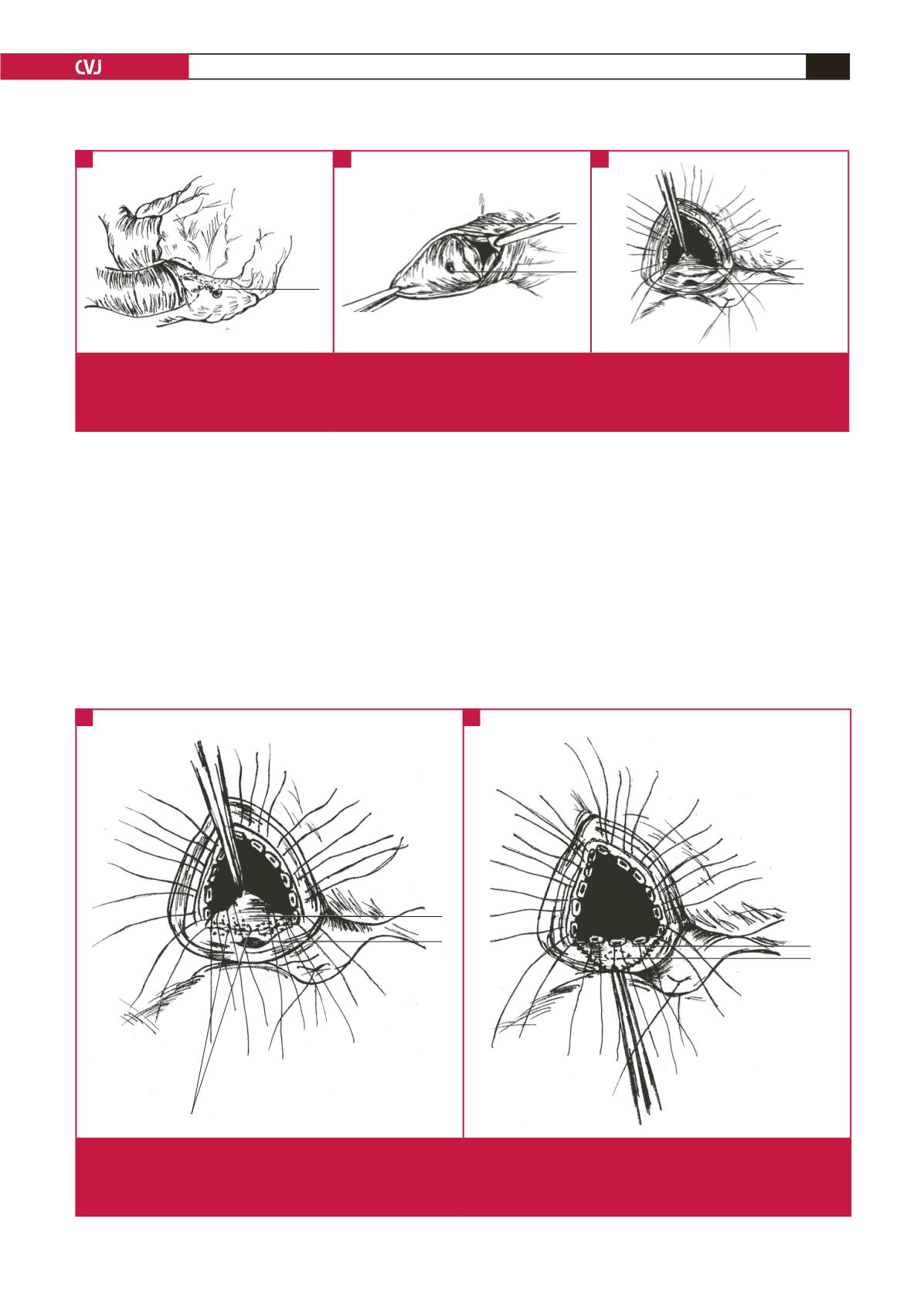
Fig. 1.
(A) Rupture of the non-coronary aneurysm of the sinus of Valsalva was revealed with the typical narrow band leading to the
right atrium. (B) 1, aorta, 2, rupture of the aneurysm of the non-coronary sinus of Valsalva. (C) The left leaflet was used to
close the narrow band of aneurysm from inside the aorta. 1, non-coronary leaflet of the aortic valve. 2, rupture of the aneu-
rysm of the non-coronary sinus of Valsalva.
A
B
C
Fig. 2.
Placement of the sutures in closing the non-coronary sinus of Valsalva with a native leaflet. (A). The left leaflet was used to
close the narrow band of the aneurysm from inside the aorta. 1, non-coronary leaflet of the aortic valve. 2, rupture of the
aneurysm of the non-coronary sinus of Valsalva. 3, sutures with pledget. (B) 1, non-coronary leaflet of the aortic valve sewn
to the aorta. 2, closed aneurysm of the non-coronary sinus of Valsalva.
A
B
1
1
1
2
2
1
2
1
2
3


