


 27 / 78

CARDIOVASCULAR JOURNAL OF AFRICA • Volume 26, No 4, July/August 2015
AFRICA
173
For the semi-quantitative assessment, the total counts for
the region of interest (ROI) were obtained on an anterior static
image (slice 15). This ROI was manually drawn (six pixels wide).
The same ROI was copied and pasted to the infra-cardiac area
below the inferior wall of the left ventricle. On the same raw
data, the images were rotated to a lateral view (slice 45), and
the ROI was copied and pasted to the inferior wall and the
corresponding infra-cardiac area (Figs 2, 3). Regions of interest
were copied between stress and rest studies of individual patients
to increase reproducibility.
Statistical analysis
Data were analysed using a Statistica 10 package (statsoft Inc,
Tilsa, Oklahoma, USA).
18
Descriptive results were presented
as medians and range (normal or not normally distributed) for
continuous variables. Categorical variables were summarised as
frequencies and percentages. To assess the differences between
continuous variables (age, counts in the left ventricle and infra-
cardiac region at rest and stress) (not normally distributed), a
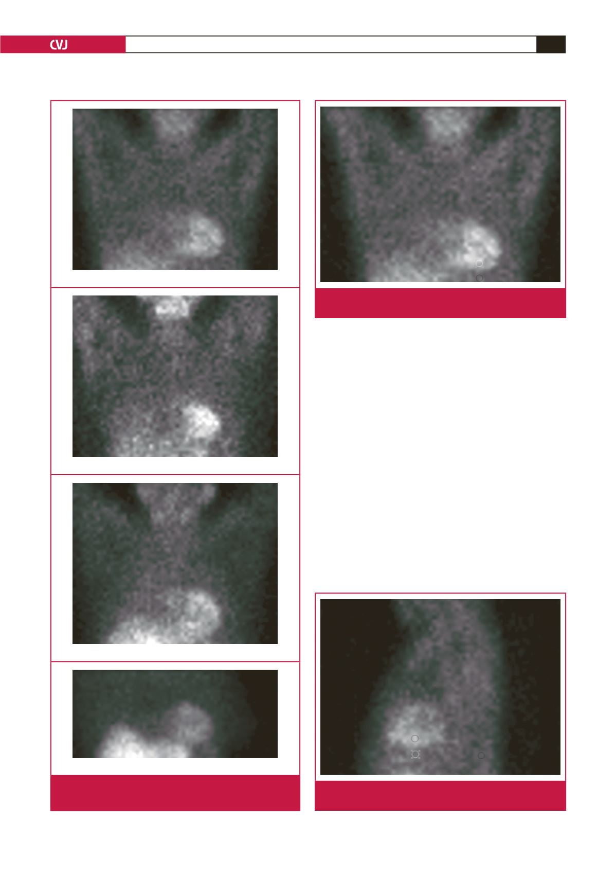
Grade 0: Absence of infra-cardiac activity
Grade 1: Infra-cardiac < myocardial activity
Grade 2: Infra-cardiac = myocardial activity
Grade 3: Infra-cardiac > myocardial activity
Fig. 1.
Example of the grading of the relative intensity of
infra-cardiac activity compared to myocardial activity
Fig. 2.
Anterior image. ROI in the inferior wall of the left ventri-
cle copied to the infra-cardiac ROI (Slice 15)
Fig. 3.
Lateral image. ROI in the inferior wall of the left ventri-
cle copied to the infra-cardiac ROI (Slice 45)