
CARDIOVASCULAR JOURNAL OF AFRICA • Volume 27, No 2, March/April 2016
AFRICA
123
Case Report
Pregnancy and childbirth in a patient after multistep
surgery and endovascular treatment of cardiovascular
disease
Piotr Buczkowski, Mateusz Pu
ś
lecki, Sebastian Stefaniak, Jerzy Kulesza, Olga Trojnarska, Tomasz
Urbanowicz, Marek Jemielity
Abstract
Nowadays physicians see an increasing population of patients
reaching reproductive age after surgery for complex congeni-
tal heart defects. Correction of congenital and acquired
cardiovascular defects does not exclude experiencing a safe
pregnancy. We present the case of a 27-year-old woman, who,
after multistep surgery and endovascular treatment of her
cardiovascular system, underwent successful pregnancy and
uncomplicated childbirth. Recent developments in medicine
and interdisciplinary involvement have allowed women with
corrected cardiovascular disease the opportunity to become
pregnant and experience safe childbirth.
Keywords:
pregnancy, childbirth, aortic aneurysm, congenital
disease, coarctation, hybrid treatment
Submitted 20/1/15, accepted 14/11/15
Cardiovasc J Afr
2016;
27
: e1–e2
www.cvja.co.zaDOI: 10.5830/CVJA-2015-084
In the past, girls with complex congenital or acquired heart
defects often did not reach reproductive age. Recent developments
in intensive paediatric cardiac surgery mean that more girls reach
the age of maturity. Interdisciplinary involvement has allowed
women with corrected cardiovascular defects the opportunity to
become pregnant and experience safe childbirth.
Case report
A 27 year-old woman in good physical condition was admitted
to the operating room of the Department of Cardiac Surgery
because of a planned pregnancy. When she was six years old, she
was operated on for a defect in the interventricular septum. After
10 months, she underwent surgical correction of aortic coarctation
using a Dacron patch. During childhood and adolescence, the
patient was normotensive and without any cardiac disease.
At the age of 22 years, new symptoms appeared in the form of
hoarseness and periodic aphonia, which suggested the presence
of a rare postoperative complication, aortic dilatation on the
border of the aortic arch and descending aorta. This suspicion
was confirmed by imaging with computed angiography, which
demonstrated dilatation of the distal aortic arch and descending
aorta, starting at a maximum of 65 to 70 mm, with a normal-
diameter (19 mm) descending aorta.
She was involved in two-stage hybrid treatment.
1
Initially, via
median sternotomy, anastomosis was performed between the
ascending aorta, brachiocephalic trunk and left common carotid
artery, using a bifurcated graft (FlowNit Bioseal 12 mm). Due
to extensive collateral circulation, the left subclavian artery was
not revascularised.
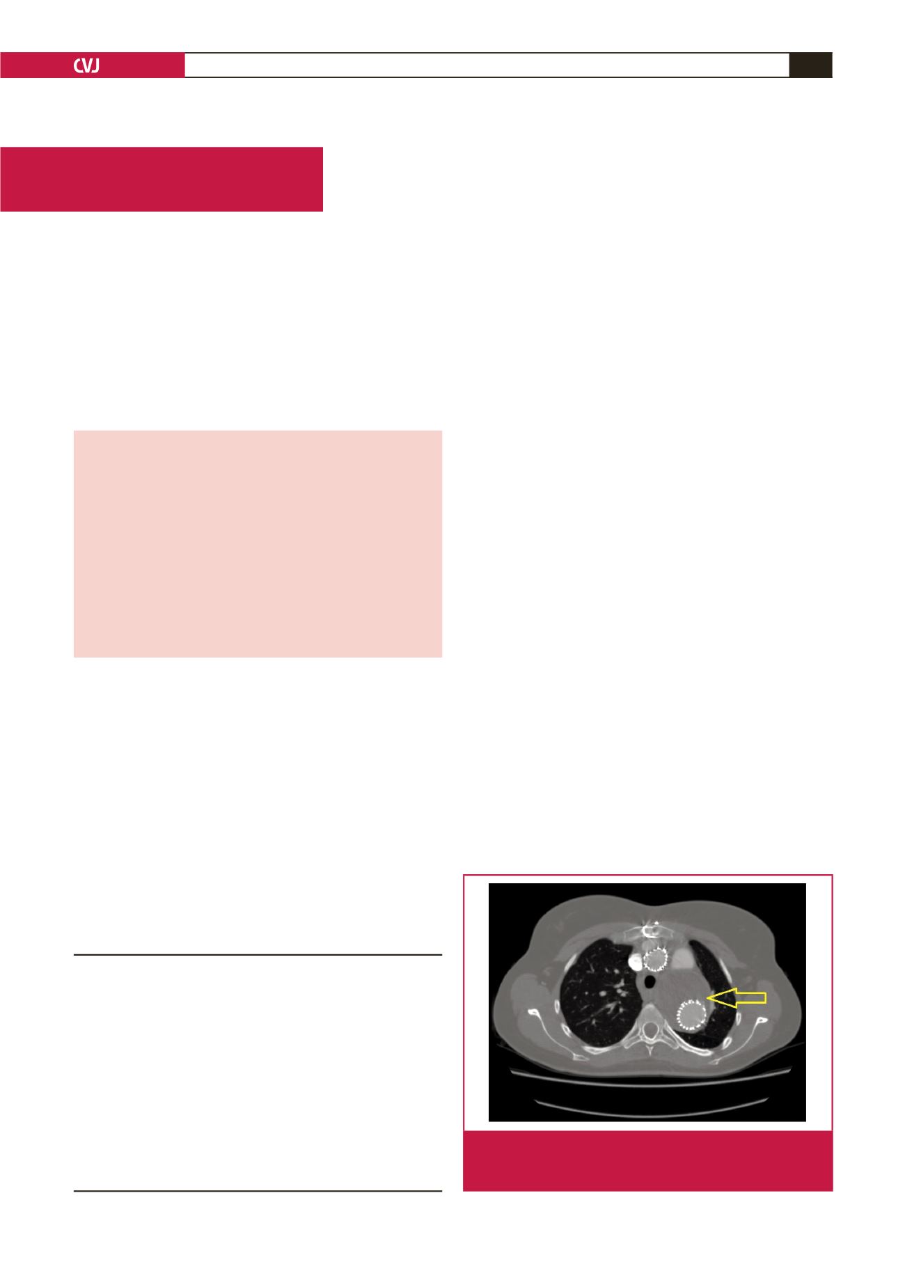
After 10 days, the second endovascular step was executed. Two
stent grafts (Zenith TX2 TAA 28 mm) were implanted in the aorta
Department of Cardiac Surgery and Transplantology,
Poznan University of Medical Sciences, Poznan, Poland
Piotr Buczkowski, MD, PhD
Mateusz Pu
ś
lecki, MD, PhD
Sebastian Stefaniak, MD, PhD,
seb.kos@gmail.comTomasz Urbanowicz, MD, PhD
Marek Jemielity, MD
Department of Radiology, Poznan University of Medical
Sciences, Poznan, Poland
Jerzy Kulesza, MD
First Department of Cardiology, Poznan University of
Medical Sciences, Poznan, Poland
Olga Trojnarska, MD
Fig. 1.
Computed tomography angiography three days after
surgery. The aneurysm sac was excluded from the
circulatory system after thoracic stent graft implantation.


