


 81 / 92

CARDIOVASCULAR JOURNAL OF AFRICA • Volume 27, No 4, July/August 2016
AFRICA
e11
A diagnosis of combined giant aneurysms of the CS and
SVC accompanied by right heart failure was made. Due to the
two giant venous aneurysms, cardiac thrombosis and severe
right heart failure, cardiac transplantation was considered the
only viable option. At the time of publication of this report, the
patient was being maintained on aspirin and furosemide while
awaiting cardiac transplantation.
Discussion
Venous aneurysms are uncommon, particularly those involving
the CS or SVC. The aetiologies of venous aneurysms are still
under debate. The causes of acquired venous aneurysms may
include trauma, inflammation or pathological processes that
affect the vascular wall.
4
Longstanding venous hypertension
secondary to heart failure, tricuspid valve lesions, cardiomyopathy
and constrictive pericarditis have also been considered to cause
vascular damage.
5
Mahmud and colleagues found that CS size was positively
correlated with right atrial size and pressure.
6
Dilatation of
the vena cava may be observed in right heart failure. Wells
et al
. reported a massive aneurysm of the IVC, and the
patient’s longstanding right heart failure was presumed to be the
underlying cause.
7
We report a case of combined giant venous aneurysms,
diagnosed using multiple imaging modalities, which provided
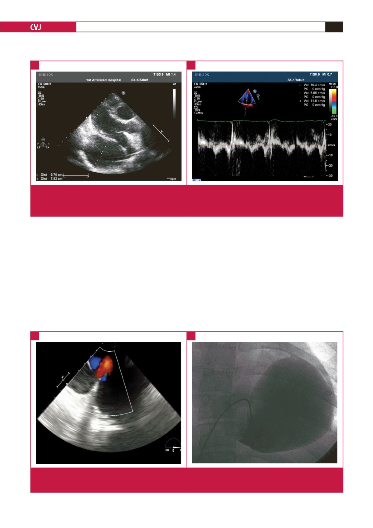
RV
LV
CSA
LA
AO
s
′
e
′
a
′
Fig. 1.
Transthoracic echocardiography on presentation showed: A. the appearance of the giant aneurysm of the coronary sinus in
the parasternal long-axis view; B. tissue Doppler imaging velocities at the septal mitral annulus: early diastolic myocardial
velocity (e
′
) was 5.85 cm/s and systolic myocardial velocity (s
′
) was 10.4 cm/s. RV, right ventricle; LA, left atrium; LV, left
ventricle; CSA, coronary sinus aneurysm; AO, ascending artoa, a
′
, late diastolic myocardial velocity.
A
B
RA
IVS
CSA
CSA
Fig. 2.
A.Transoesophageal echocardiography colour Doppler flow imaging demonstrated flow from the coronary sinus aneurysm to
the right atrium. B. Cardiac catheterisation showed the appearance of the giant coronary sinus aneurysm. RA, right atrium;
IVC, inferior vena cava; CSA, coronary sinus aneurysm.
A
B