


 82 / 92

CARDIOVASCULAR JOURNAL OF AFRICA • Volume 27, No 4, July/August 2016
e12
AFRICA
strong evidence for an aetiology of longstanding right heart
diastolic failure.
Aneurysm of the CS is a rare abnormality of the intracardiac
vein.
8
Previous cases in the literature were considered to be
congenital or secondary to anomalous drainage.
9,10
We excluded
the possibility of congenital causes on the basis of previous CT
examinations. Echocardiography and contrast-enhanced CT gave
a precise anatomical view of the CS and SVC, demonstrating the
absence of anomalous drainage.
Constrictive pericarditis (CP) is pathologically characterised
by scarring and a loss of pericardium elasticity, resulting in
an external interruption of cardiac filling.
11
Pericardiectomy
remains the most effective therapy for CP. Diastolic dysfunction
and low-output syndrome occur in a considerable number
of patients after pericardiectomy, which may be the result
of atrophic changes in the myocardium associated with
longstanding pericardial restriction.
12
Our patient had a long
history of CP before pericardiectomy. On the basis of previous
echocardiography, we concluded that the patient had had a long
history of right heart diastolic failure.
Echocardiography also revealed the progression of combined
venous aneurysms. Colour Doppler echocardiography revealed
to-and-fro flow between the CS aneurysm and right atrium.
Catheter examination demonstrated equally increased pressure in
*
**
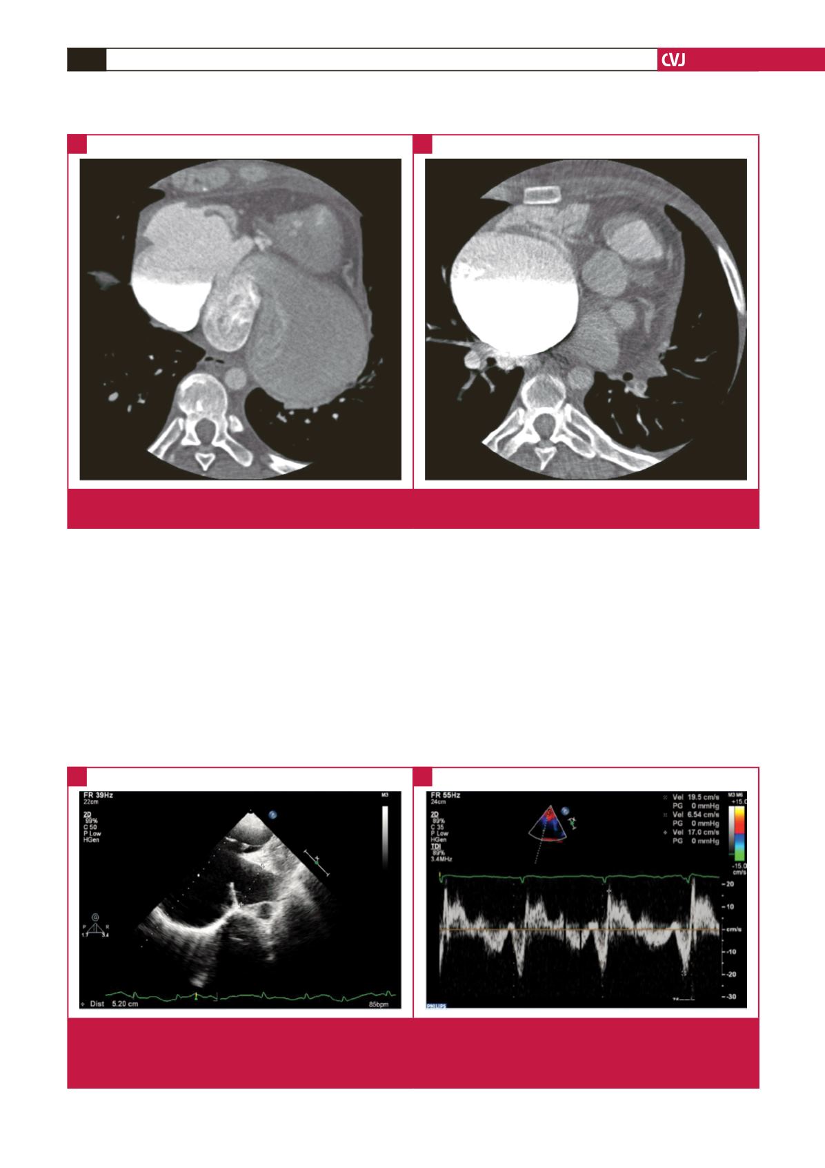
Fig. 3.
Contrast-enhanced CT showed a giant aneurysm of the coronary sinus ( * ) and a giant aneurysm of the superior vena cava
( ** ).
A
B
RV
RA
CSA
s
′
e
′
a
′
Fig. 4.
Transthoracic echocardiography performed four years prior to presentation showed: A. the aneurysmal dilated CS was 5 cm
in diameter; B. tissue Doppler imaging velocities at the septal mitral annulus: early diastolic myocardial velocity (e
′
) was 6.54
cm/s and systolic myocardial velocity (s
′
) was 19.5 cm/s. RV, right ventricle; RA, right atrium; CSA, coronary sinus aneurysm;
a
′
, late diastolic myocardial velocity.
A
B