
CARDIOVASCULAR JOURNAL OF AFRICA • Volume 27, No 6, November/December 2016
AFRICA
347
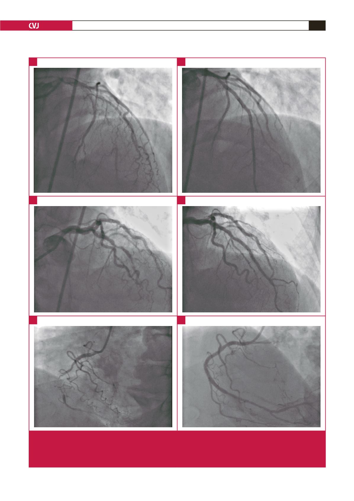
Fig. 1.
Coronary angiography showing recanalisation of chronic total occlusions. (A) Chronic total occlusion of the LAD from the
proximal portion. (B) Successfull recanalisation of the LAD with two BVSs (3.5 × 28 mm and 3.0 × 28 mm) via antegrade
approach. (C) Chronic total occlusion of the LAD from the mid portion. (D) Successfull recanalisation of LAD with two BVSs
(3.5 × 18 mm and 3.0 × 28 mm ) via antegrade approach. (E) Chronic total occlusion of the RCA from the mid portion. (F)
Successfull recanalisation of the RCA with one BVS (3.0 × 18 mm) via antegrade approach with the aid of a microcatheter.
A
C
E
B
D
F


