


 8 / 88

CARDIOVASCULAR JOURNAL OF AFRICA • Volume 28, No 4, July/August 2017
210
AFRICA
Echocardiographic images of EMF were divided into
the following subgroups: images of basic echocardiographic
features (covered in Figs 1–3), images of additional
echocardiographic details (shown in Fig. 4) and images of
new echocardiographic features (shown and described in Figs
5 and 6).
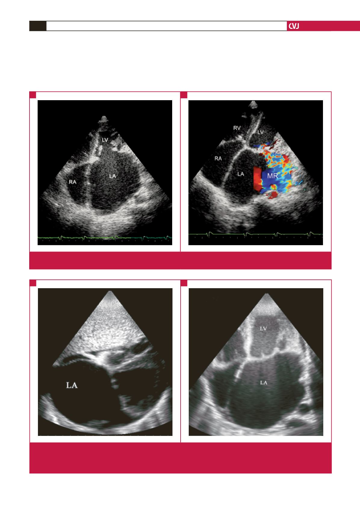
Fig. 1.
Basic echocardiographic features of left ventricular EMF. A and B, apical four-chamber view. The huge left atrium with apical
and LV wall fibrosis, obliterated LV and mitral regurgitation (MR) (B), are the characteristic features of left ventricular EMF.
A
B
Fig. 2.
Basic echocardiographic features of advanced left ventricular EMF. Images from a 42-year-old female who presented with
intractable heart failure. A is a subcostal view showing the typically huge left atrium occupying half the cardiac size. B is an
AP four-chamber view showing apical fibrosis extending to the septum and ventricular walls, leading to severe obliteration
of the left ventricular cavity.
A
B