


 9 / 88

CARDIOVASCULAR JOURNAL OF AFRICA • Volume 28, No 4, July/August 2017
AFRICA
211
Discussion
This descriptive study that discusses the clinical and
echocardiographic features of EMF in Sudan is not intended to
redefine the pathognomonic echocardiographic features of the
disease, which have been well described in previous works.
3,4,6,22
The main objective was to determine the current frequency of
EMF in Sudan, as the last report on the disease dates back more
than 40 years.
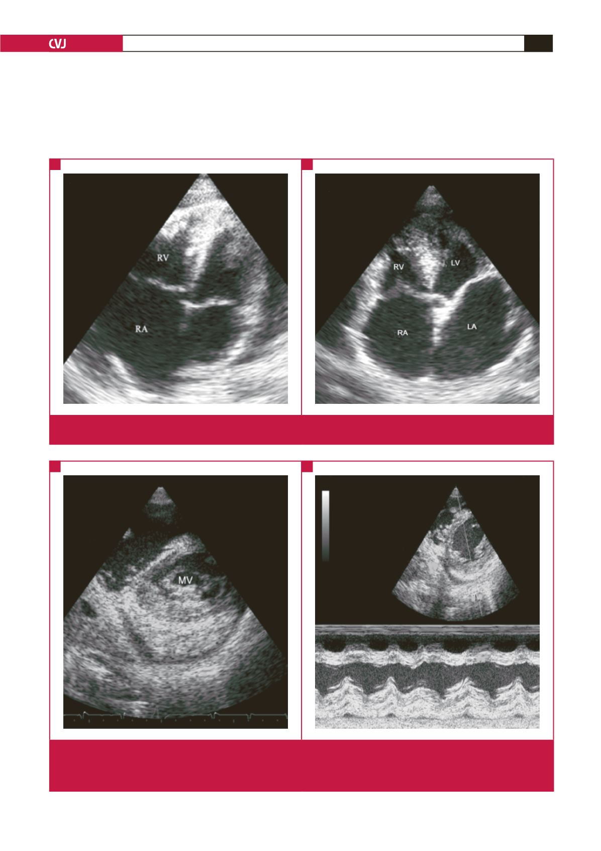
Fig. 3.
Right ventricular (RV) and biventricular EMF. A (AP4) showing EMF of the RV; note the apical fibrosis engulfing the modera-
tor band, fibrosis of the anterior interventricular septum, dilated RA and obliterated RV. B (AP5) shows biventricular EMF.
A
B
Fig. 4.
Layering. A is a short-axis view of a patient with advanced EMF showing layering of the thickened endocardium with the
myocardium and pericardium. The posterior mitral valve leaflet (MV) is seen tethered to the endocardium. B is M-mode
showing the distinct layers of the posterior wall with thickened endocardium and myocardium. A thickened pericardium with
effusions can be seen peripherally.
A
B