


 78 / 82

CARDIOVASCULAR JOURNAL OF AFRICA • Volume 29, No 1, January/February 2018
e12
AFRICA
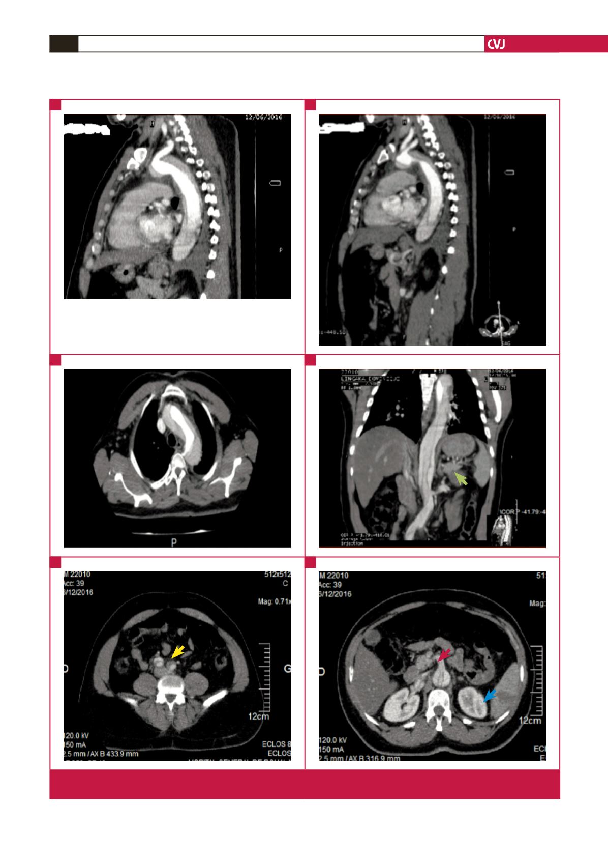
Fig. 4.
Contrast-enhanced CT angiogram of the thorax showing aortic dissection extending to the left renal (green arrow), iliac
(yellow arrow) and superior mesenteric (red arrow) arteries and causing splenic infarction (blue arrow).
A
C
E
B
D
F