


 27 / 84

CARDIOVASCULAR JOURNAL OF AFRICA • Volume 29, No 2, March/April 2018
AFRICA
89
The majority of embolic occlusions occurred at the level of the
brachial artery bifurcation (
n
=
17). Nine patients presented
with more proximal (two subclavian artery, seven axillary
artery) and three with more distal occlusions (two radial artery,
one ulnar artery). One patient presented with a blue-finger
syndrome. A left-sided predominance was noted across all levels
of obstruction with a right-to-left-sided ratio of 1:2.
A distinct proximal distribution of atherosclerotic lesions was
observed, with the subclavian artery involved in eight, the axillary
artery in one and the brachial artery in three patients. As observed
in the thrombo-embolic subgroup, clear left-sided predominance
was noted with a right-to-left ratio of 1:5. Morphologically, six
lesions were described as stenotic and five as occlusions.
Arterial thoracic outlet syndrome: seven of eight patients
presented with underlying bony pathology (five cervical ribs, one
anomalous first rib and one old clavicle fracture resulting in a
pseudo-arthrosis). Six patients presented with chronic and two
with acute ULI.
Four patients were diagnosed with Takayasu’s disease. Three
patients presented with upper-limb claudication. One of these
claudicants suffered an ipsilateral ischaemic cerebrovascular
accident prior to presentation. Level of disease ranged from
stenosis of the innominate artery with occlusion of its outflow
(one patient) to proximal left common carotid artery stenosis
with associated left subclavian artery occlusion (two patients).
One patient presented with prosthetic graft sepsis complicated
by an acute anastomotic bleed following previous aortic arch
reconstruction for aneurysmal disease.
Thrombo-angiitis obliterans: four patients with active digital
ulceration due to Buerger’s disease were evaluated and surgically
managed during the study period. The average smoking history
was 36 pack years.
Small-vessel disease: two patients presented with active
digital ulceration in combination with Reynaud’s symptoms and
one presented with Reynaud’s symptoms alone. The vascular
pathologies identified were a vasculitis (Lupus), a hypothenar
hammer syndrome and an atherosclerotic small-vessel disease.
Clinical presentation
Thirty-five patients (54.7%) presented with acute ULI
necessitating surgical intervention. Three patients (8.6%) had
signs of irreversible ischaemia (Rutherford grade III ULI) and
a further nine (25.7%) were diagnosed with Rutherford grade
IIb ULI.
Twenty-nine patients presented with chronic ULI. Fourteen
patients (48.3%) presented with tissue loss. Other clinical
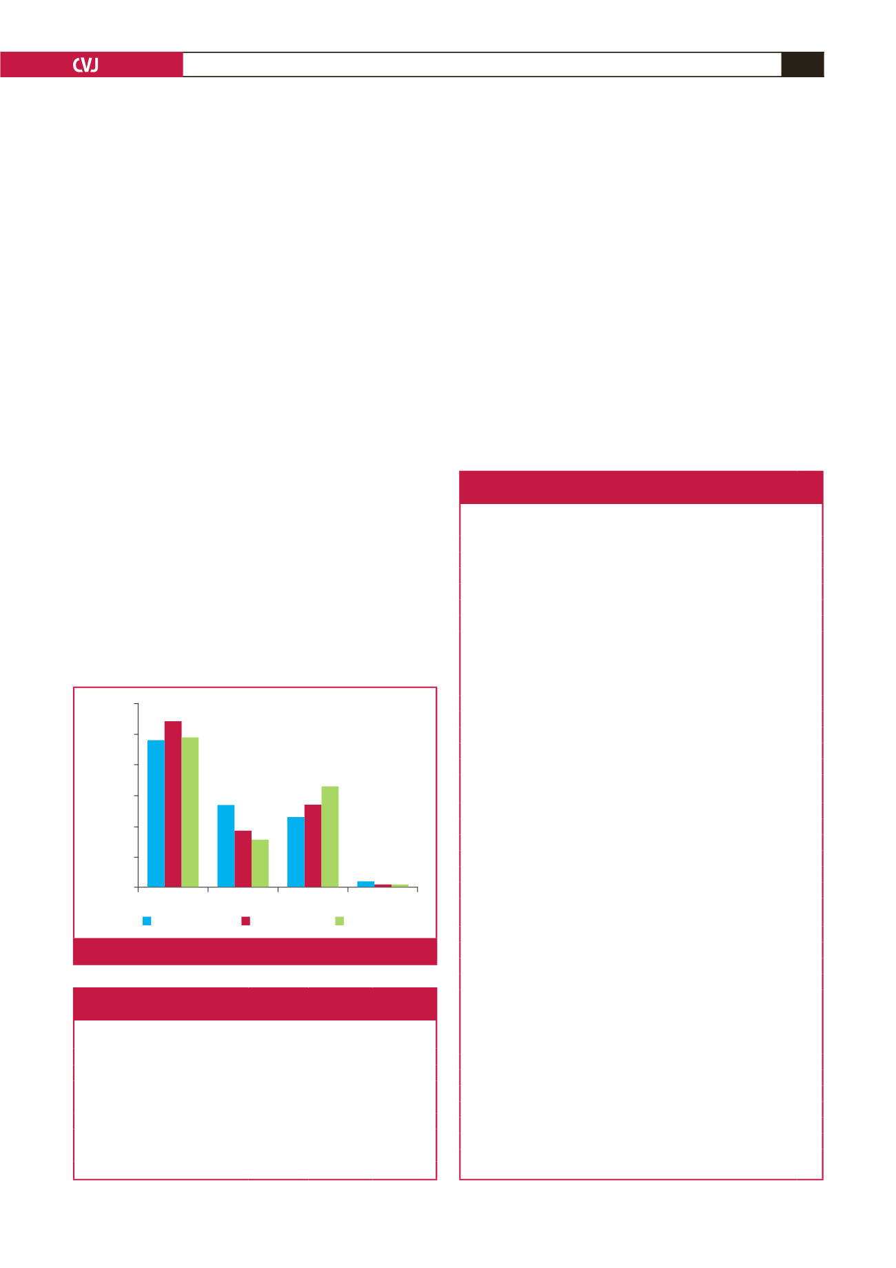
Mixed-race Caucasian
Black
Indian
Percentage
60
50
40
30
20
10
0
Current study
Census 2001
Census 2011
Fig. 1.
Racial prevalence.
Table 1. Comparative demographic details
according to vascular pathology
Pathology
Number of
patients
Mean age
(range)
Male:female
ratio
Thrombo-embolic disease
30
55 (37–80)
0.43
Atherosclerotic disease
12
57 (39–84)
0.71
Thoracic outlet syndrome
8
28 (15–59)
1.67
Takayasu’s disease
4
27 (20–36)
0.33
Thrombo-angiitis obliterans
4
46 (36–53)
3.00
Small-vessel disease
3
32 (31–49)
0.50
Iatrogenic
2
–
1.00
Polymyositis compartment syndrome
1
–
–
Table 2. Procedures performed according to aetiology
(excluding minor and ablative procedures)
Procedures performed
No.
Thrombo-embolic disease
35
Thrombo-embolectomy (fasciotomy in eight)
25
Brachial–brachial/–distal bypass (autologous vein graft)
4
Catheter-directed/intra-operative thrombolysis
3
Stent–graft placement
1
Aortic arch reconstruction (Gelsoft
®
Dacron)
1
Veinpatch angioplasty of veingraft
1
Atherosclerotic occlusive disease
19
Subclavian artery stent placement
5
Brachial–distal/brachial–brachial bypass graft
4
Subclavian–axillary/subclavian–brachial bypass graft
3
Axillary–brachial bypass graft
2
Common carotid–axillary/common carotid–brachial bypass graft
2
Graft thrombectomy
2
Subclavian artery balloon angioplasty
1
ATOS (each row represents a patient)
11
Thrombo-embolectomy + fasciotomy; Ipsilateral TO decompression;
contralateral TO decompression
3
Thrombo-embolectomy + fasciotomy; Ipsilateral TO decompression
2
Common carotid–brachial RSBG bypass
1
Subclavian–axillary PTFE bypass + brachial–ulnar RSVG bypass
1
Subclavian–axillary PTFE bypass
1
Ipsilateral TO decompression
1
Subclavian–axillary PTFE bypass
1
Common carotid–brachial Dacron
®
bypass + brachial–ulnar RSVG
bypass + fasciotomy
1
Takayasu’s disease
4
Aortic arch reconstruction AlboGraft
®
2
Redo aortic arch reconstruction SilverGraft
®
1
Axillary–axillary PTFE bypass
1
Thrombo-angiitis obliterans
2
Thoracoscopic sympathectomy
2
Small-vessel disease
3
Thoracoscopic sympathectomy
2
Brachial–distal RSVG bypass
1
Iatrogenic (each row represents a patient)
2
Fasciotomy
1
Thrombo-embolectomy + fasciotomy
1
Polymyositis compartment syndrome
1
Fasciotomy
1
TO: thoracic outlet; RBVG: reverse basilic vein graft; RSVG: reverse saphenous
vein graft; PTFE: polytetrafluoroethylene.